


'/%3e%3c/svg%3e)

- By Indication
 Inflammatory and Autoimmune DiseasesRespiratory DiseasesInfectious DiseasesRare DiseaseGut Microbiota Research ServiceBy Modality
 Preclinical Model Strategies for T-Cell Engagers: From Oncology to Autoimmune Disease
 2026-10-27
 11:00 AM ET
 Dr. Wanjun Yuan & Dr. Haiqiao SunOne TCE Modality. Two Disease Contexts. Different Preclinical Questions.

'/%3e%3c/svg%3e)
'/%3e%3c/svg%3e)

Metabolic
Translational Endometriosis-Associated Pain Models for Evaluating Novel Therapies
Endometriosis affects millions of women worldwide with substantial unmet clinical needs. GemPharmatech's translational mouse and rat models offer a clinically relevant platform for evaluating novel therapies in this underserved area of women's health.
 2026-08-26

Endometriosis (EMT) is a chronic gynecological disorder characterized by the presence and growth of endometrial-like tissue outside the uterine cavity. Ectopic lesions commonly occur in pelvic organs and peritoneal tissues, resulting in repeated bleeding, inflammation, fibrosis, and pain.
Endometriosis affects approximately 10% of women of reproductive age worldwide and is strongly associated with infertility and chronic pelvic pain[1]. Pain-related symptoms, including dysmenorrhea, chronic pelvic pain, and deep dyspareunia, represent major clinical burdens.
Endometriosis-associated pain is driven by a complex interaction among hormonal regulation, inflammatory responses, and nervous system sensitization.
Estrogen-dependent ectopic lesion growth contributes to local tissue remodeling and activation of peripheral nociceptive pathways. Persistent stimulation from ectopic lesions may further promote neuronal sensitization and abnormal pain signaling.
Increasing evidence suggests that neurogenic mechanisms, including altered nerve growth and activation of pain-related pathways such as NGF/BDNF/TRPV1 signaling, play important roles in the development and maintenance of endometriosis-associated pain[5-7].
1. Endometriosis-Associated Pain: A Complex Neuroimmune Process
Endometriosis-associated pain is driven by the interaction of hormonal regulation, immune inflammation, and nervous system sensitization[2-4].
Estrogen-dependent lesion growth promotes inflammatory responses and activation of peripheral nociceptors. Meanwhile, ectopic lesions recruit immune cells and increase secretion of inflammatory mediators including IL-1β, IL-6, TNF-α, and PGE2[4], leading to pain hypersensitivity.
Long-term inflammatory stimulation induces abnormal nerve growth and sensitization pathways involving NGF, BDNF, and TRPV1, contributing to persistent pain[5-7].
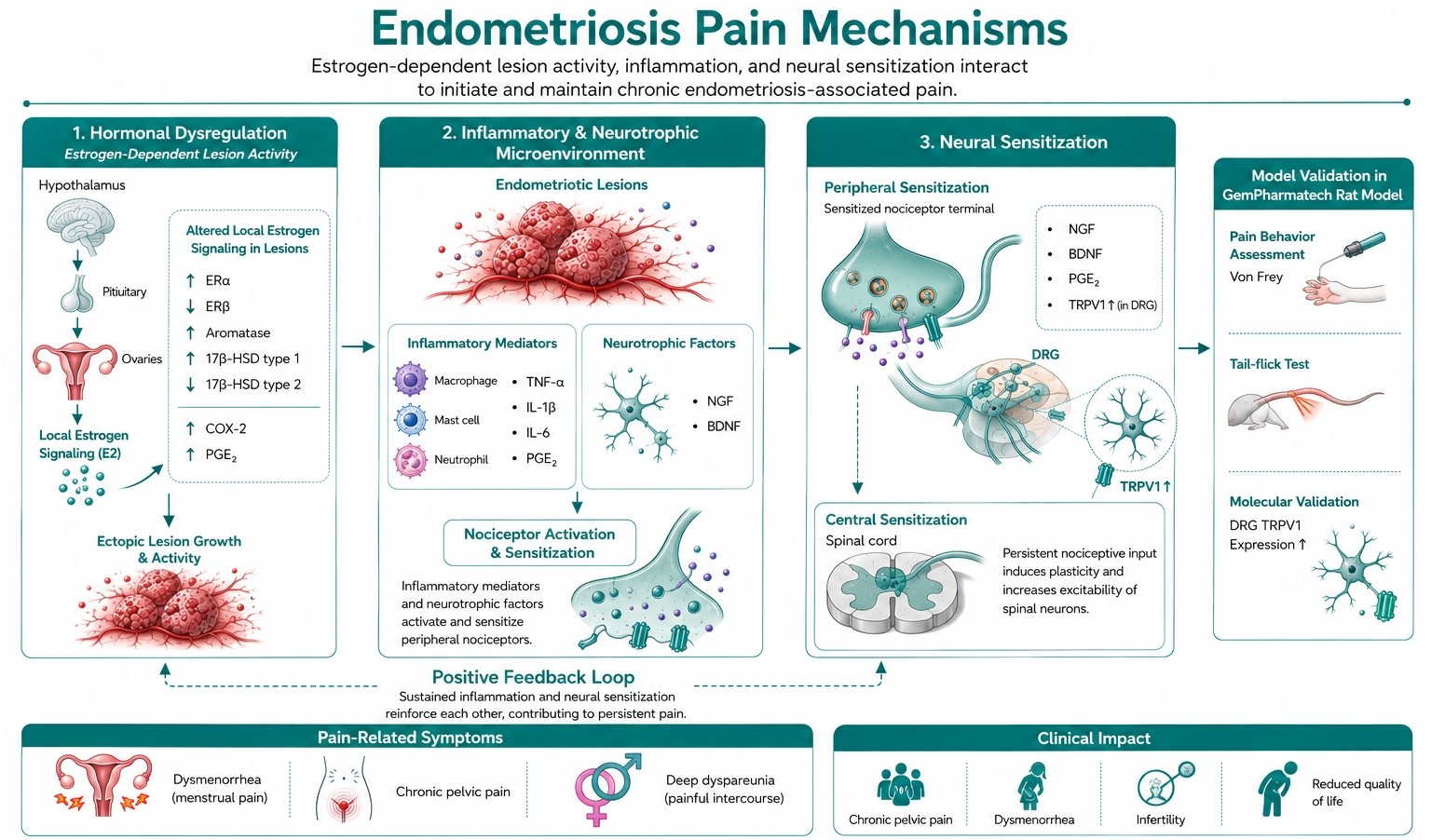
Figure 1. Mechanisms of Endometriosis-Associated Pain
2. Establishment of Rat Endometriosis Pain Model
To establish a clinically relevant rat endometriosis model, female SD rats were divided into three groups: sham-operated controls, EMT model rats undergoing surgery during the natural estrus phase, and estrogen-supported EMT model rats with sustained hormonal stimulation.
Endometrial tissue fragments were collected, standardized, and transplanted onto the abdominal wall[8] with the endometrial surface exposed to facilitate ectopic lesion formation. For the estrogen-supported EMT group, estrus was induced before surgery and maintained through repeated estrogen administration (Q2W) after surgery, enhancing lesion establishment and disease progression. Sham-operated animals underwent the same surgical procedure with adipose tissue transplantation instead of endometrial tissue.
The model design enables evaluation of ectopic lesion establishment, lesion progression, pathological changes, and pain-related behavioral responses.
Autologous uterine tissue is transplanted onto the abdominal walI during the estrus phase. Lesion formation and pain phenotypes are evaluated by behavioral tests and molecular analyses.
3. Successful Establishment of Endometriotic Lesions
Model validation demonstrated successful formation of ectopic endometrial lesions.
Gross examination confirmed the successful establishment of ectopic endometrial lesions, which presented as typical cyst-like structures with visible vascularization at Day 28 after transplantation. Histological evaluation by hematoxylin and eosin (H&E) staining further confirmed the presence of ectopic endometrial tissues, characterized by preserved epithelial components, glandular structures, stromal proliferation, and inflammatory cell infiltration.
Both the EMT Model and EMT+Estradiol Model exhibited well-developed ectopic lesions with characteristic endometrial morphology, confirming that both approaches successfully established the rat endometriosis (EMT) model[8].
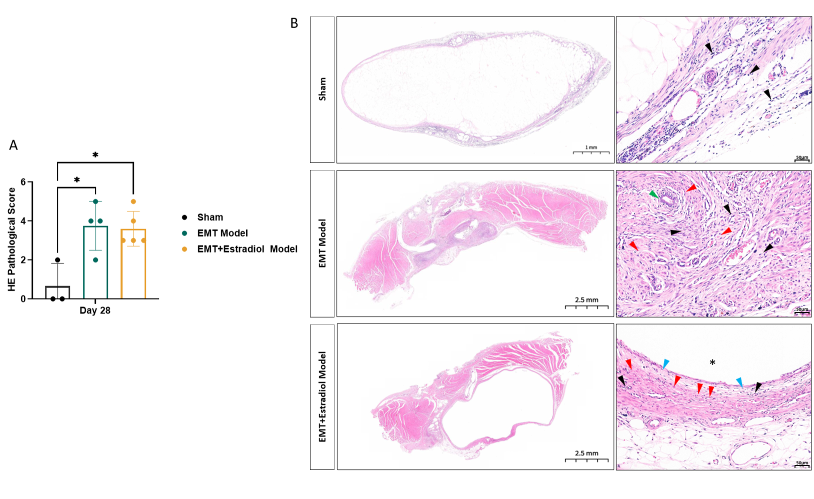
Figure 2. Histopathological validation of the rat endometriosis (EMT) model by H&E staining.
(A) Histopathological scores of ectopic lesions evaluated based on epithelial integrity, glandular/cystic structure formation, stromal hyperplasia, and inflammatory cell infiltration. Data are presented as mean ± SEM (n=3-5 per group). *P < 0.05; **P < 0.01; ***P < 0.001; ****P < 0.0001 (one-way ANOVA). (B) Representative hematoxylin and eosin (H&E)-stained sections of ectopic lesions from the Sham, EMT Model, and EMT + Estradiol Model groups. No obvious ectopic endometrial structures were observed in the Sham group. The EMT Model group developed typical ectopic endometrial lesions, whereas continuous estradiol stimulation further maintained lesion development and induced more pronounced histopathological alterations in the EMT + Estradiol Model group. Blue arrowheads indicate the monolayer endometrial epithelium; green arrowheads indicate endometrial glands; red arrowheads indicate stromal cells; black arrowheads indicate inflammatory cell infiltration; asterisks (*) indicate the uterine lumen. H&E stain; Scale bars: 1/2.5 mm (low-magnification images) and 50 μm (high-magnification images).
4. Pain Phenotyping and Validation
To evaluate endometriosis-associated pain, multiple behavioral assays were incorporated.
Mechanical hypersensitivity was assessed using Von Frey filament testing[9], including evaluation of lesion-associated abdominal pain and peripheral hypersensitivity.
Thermal nociception was evaluated using the tail-flick assay[10] to assess heat sensitivity changes during disease progression.
The model showed reproducible pain-related phenotypes, including increased mechanical sensitivity and altered thermal pain responses after model induction.
Mechanical withdrawal thresholds were measured by applying calibrated Von Frey filaments to the plantar surface of the hind paw through an elevated wire mesh floor.
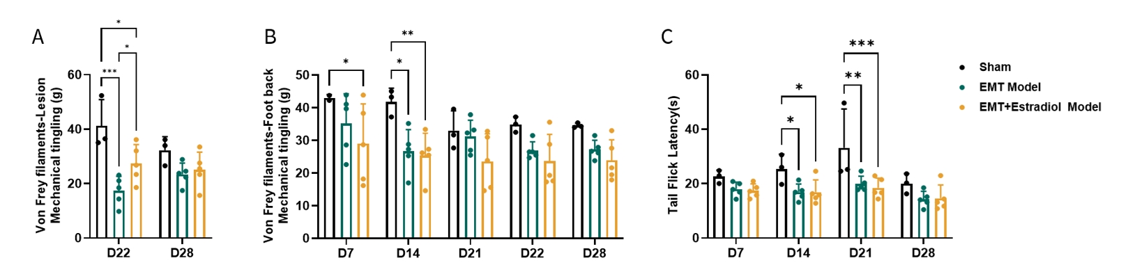
Figure 3. Behavioral assessment of endometriosis-associated pain in the rat EMT model.
(A) Mechanical sensitivity of ectopic lesions evaluated using the Von Frey filament test on Days 22 and 28. (B) Mechanical withdrawal threshold of the hind paw measured using the Von Frey filament test on Days 7, 14, 21, 22, and 28. (C) Thermal nociceptive sensitivity assessed using the tail-flick test on Days 7, 14, 21, and 28. Each point represents the average of 3 measurements taken by an animal. Data are presented as mean ± SEM. *P < 0.05; **P < 0.01; ***P < 0.001; ****P < 0.0001 (one-way ANOVA).
5. Molecular Validation of Pain Pathways
In addition to behavioral assessment, molecular characterization was performed to evaluate pain-related mechanisms.
TRPV1 expression analysis in dorsal root ganglia (DRG) demonstrated activation of pain-associated neuronal pathways, supporting the involvement of peripheral sensitization[5-7] in the model.
Combined behavioral and molecular evidence indicates that the rat endometriosis model successfully recapitulates key features of endometriosis-associated pain.
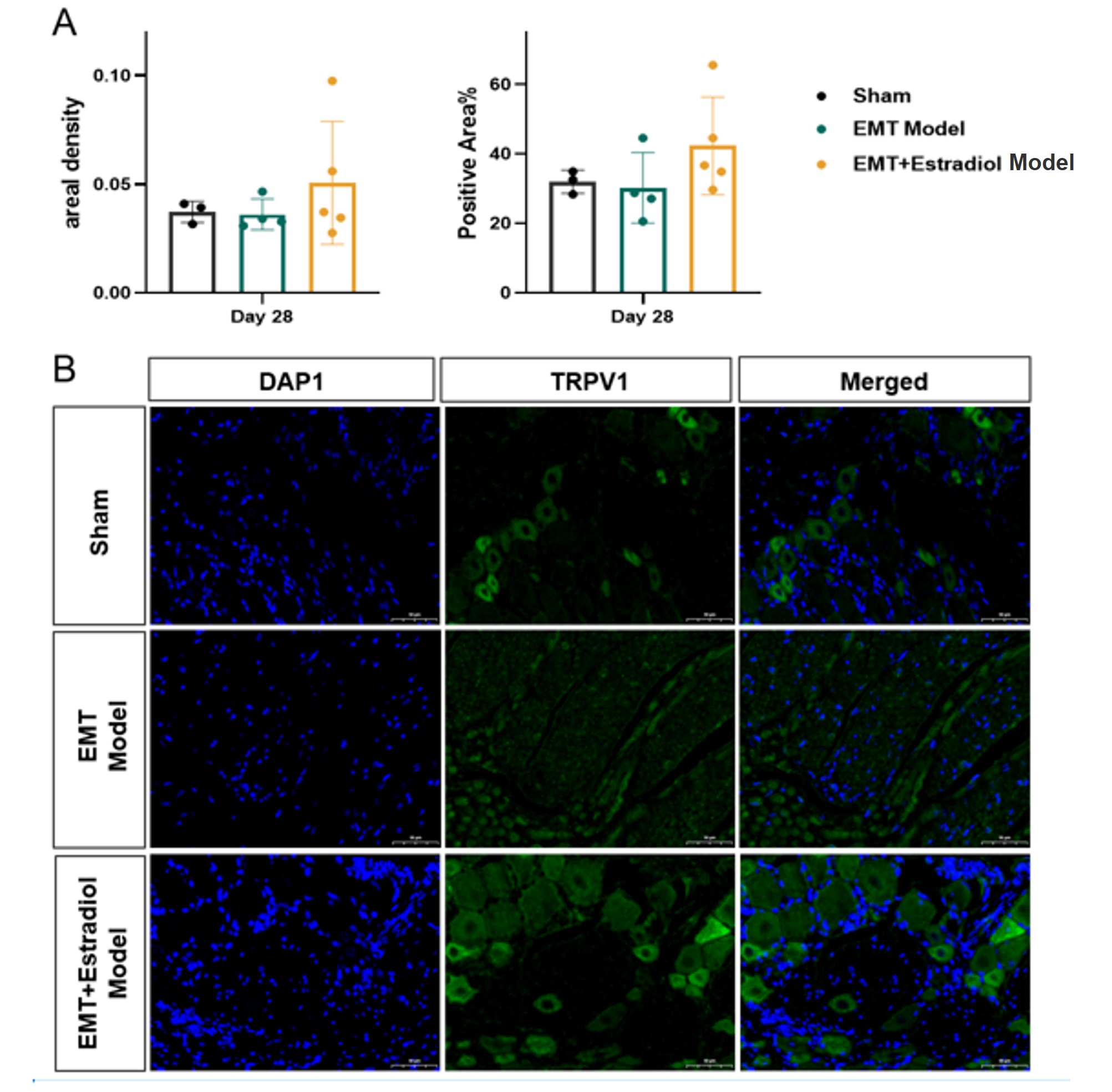
Figure 4. Effects of estradiol treatment on ectopic lesion development and TRPV1 expression in the rat endometriosis model.
(A) Quantitative analysis of ectopic lesion fluorescence at Day 28. Ectopic lesions were detected in both the EMT Model and EMT + Estradiol Model groups. Estradiol-treated animals showed a trend toward increased fluorescence-positive area and positive signal area compared with the EMT Model group; however, the differences did not reach statistical significance. (B) Representative fluorescence images of dorsal root ganglia (DRG). Green fluorescence indicates TRPV1-positive signals, and blue fluorescence indicates cell nuclei (DAPI). TRPV1 fluorescence was observed in the DRG of all groups, with a higher fluorescence intensity visually observed in the EMT + Estradiol Model group. Data are presented as mean ± SEM. Scale bars = 50 μm.
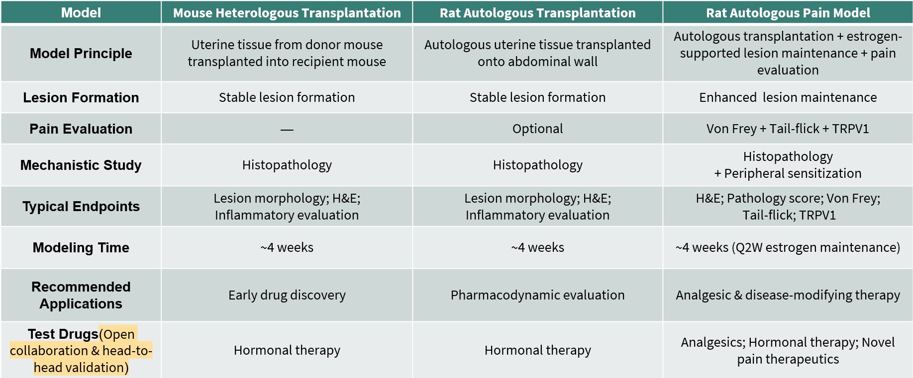
6. Endometriosis Model Platform: Comprehensive Preclinical Models for Lesion Formation and Pain Evaluation
Endometriosis is a complex chronic disease characterized by ectopic endometrial tissue growth, lesion progression, and endometriosis-associated pain. Different therapeutic strategies require disease models that accurately reproduce specific clinical phenotypes, from ectopic lesion formation to pain sensitization.
To support diverse drug discovery needs, GemPharmatech has established a comprehensive endometriosis model platform, including mouse heterologous transplantation models, rat autologous transplantation models, and rat autologous pain models.
The mouse model provides a scalable platform for early-stage drug screening, while the rat autologous transplantation model enables reliable evaluation of lesion formation and pharmacodynamic effects. Furthermore, the rat autologous pain model integrates stable ectopic lesion establishment with behavioral pain assessment and neuronal sensitization analysis, providing a translational platform for evaluating novel analgesic and disease-modifying therapies.
7. Women's Health Disease Models: Accelerating Translational Research
To address the growing demand for preclinical research in women's health and inflammatory pain disorders, GemPharmatech has established a comprehensive portfolio of disease models covering gynecological and metabolic disorders.
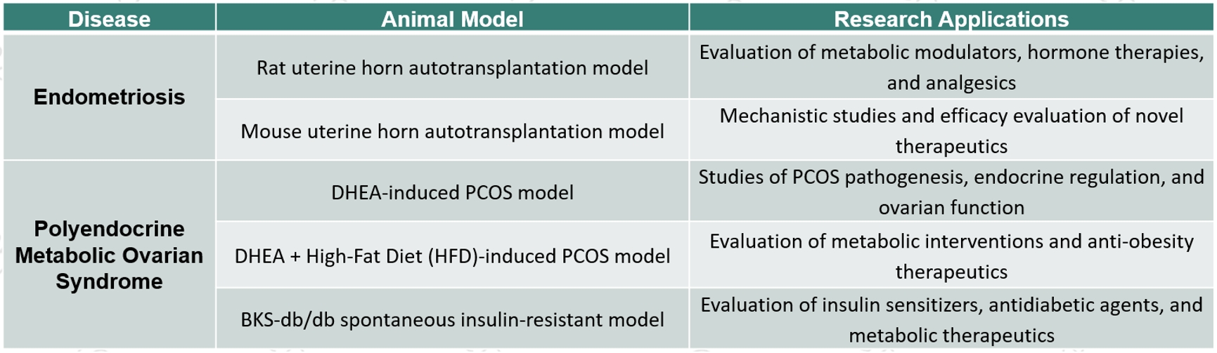
By combining well-characterized animal models with multidimensional efficacy assessment, GemPharmatech enables efficient translation from disease mechanism studies to preclinical drug development, accelerating the discovery of innovative therapies for women's health.
8. Conclusion
GemPharmatech's rat endometriosis pain model provides an integrated translational platform combining stable ectopic lesion formation, clinically relevant pain phenotyping, and mechanistic pathway validation.
With established capabilities in autologous transplantation, behavioral pain testing, histopathological evaluation, and molecular analysis, this platform supports comprehensive efficacy assessment of analgesic and disease-modifying therapies for endometriosis.
Models to Defy Impossible — Accelerating Innovation in Women's Health Research.
Reference:
1. Zondervan KT, Becker CM, Missmer SA. Endometriosis. N Engl J Med. 2020;382(13):1244–1256. doi:10.1056/NEJMra1810764.
2. Taylor HS, Kotlyar AM, Flores VA. Endometriosis is a chronic systemic disease. Nat Rev Dis Primers. 2021;7:20. doi:10.1038/s41572-021-00279-2.
3. Burney RO, Giudice LC. Pathogenesis and pathophysiology of endometriosis. Fertil Steril. 2012;98(3):511–519. doi:10.1016/j.fertnstert.2012.06.029.
4. Bulun SE. Endometriosis. N Engl J Med. 2009;360(3):268–279. doi:10.1056/NEJMra0804690.
5. Morotti M, Vincent K, Becker CM. Mechanisms of pain in endometriosis. Eur J Obstet Gynecol Reprod Biol. 2017;209:8–13. doi:10.1016/j.ejogrb.2016.07.497.
6. McAllister SL, McGinty KA, Williams ED, et al. Endometriosis-induced pain: mechanisms and therapeutic opportunities. Front Mol Neurosci. 2023;16:1132315. doi:10.3389/fnmol.2023.1132315.
7. Berkley KJ, Rapkin AJ, Papka RE. The pains of endometriosis. Science. 2005;308(5728):1587–1589. doi:10.1126/science.1111445.
8. Vernon MW, Wilson EA. Studies on the surgical induction of endometriosis in the rat. Fertil Steril. 1985;44(5):684–694.
9. Chaplan SR, Bach FW, Pogrel JW, Chung JM, Yaksh TL. Quantitative assessment of tactile allodynia in the rat paw. J Neurosci Methods. 1994;53(1):55–63. doi:10.1016/0165-0270(94)90144-9.
10. D'Amour FE, Smith DL. A method for determining loss of pain sensation. J Pharmacol Exp Ther. 1941;72:74–79.

Share


Recommended for You
More infomation about blogs

Find Models


'%3e%3cpath%20d='M13.6882%2012L23.6426%202.06059C23.8678%201.83321%2023.9941%201.52611%2023.9941%201.20607C23.9941%200.886035%2023.8678%200.578937%2023.6426%200.351553C23.4152%200.126341%2023.1081%200%2022.7881%200C22.468%200%2022.1609%200.126341%2021.9335%200.351553L11.9941%2010.306L2.05473%200.351553C1.82735%200.126341%201.52025%200%201.20021%200C0.880175%200%200.573077%200.126341%200.345693%200.351553C0.120482%200.578937%20-0.00585938%200.886035%20-0.00585938%201.20607C-0.00585937%201.52611%200.120482%201.83321%200.345693%202.06059L10.3001%2012L0.345693%2021.9394C0.120482%2022.1668%20-0.00585937%2022.4739%20-0.00585938%2022.7939C-0.00585938%2023.114%200.120482%2023.4211%200.345693%2023.6484C0.573077%2023.8737%200.880175%2024%201.20021%2024C1.52025%2024%201.82735%2023.8737%202.05473%2023.6484L11.9941%2013.694L21.9335%2023.6484C22.1666%2023.8397%2022.4624%2023.9374%2022.7635%2023.9226C23.0646%2023.9079%2023.3494%2023.7816%2023.5626%2023.5684C23.7757%2023.3553%2023.902%2023.0704%2023.9168%2022.7694C23.9316%2022.4683%2023.8338%2022.1724%2023.6426%2021.9394L13.6882%2012Z'%20fill='black'%20fill-opacity='0.96'/%3e%3c/g%3e%3cdefs%3e%3cclipPath%20id='clip0_2458_12695'%3e%3crect%20width='24'%20height='24'%20fill='white'/%3e%3c/clipPath%3e%3c/defs%3e%3c/svg%3e)
Submit Inquiry
Contact Us
Schedule a Meeting
Let us know which event you're attending and we'd be happy to connect with you.
Download
Download
Watch On-Demand Webinar
Newsletter Signup
'%3e%3cpath%20d='M13.6882%2012L23.6426%202.06059C23.8678%201.83321%2023.9941%201.52611%2023.9941%201.20607C23.9941%200.886035%2023.8678%200.578937%2023.6426%200.351553C23.4152%200.126341%2023.1081%200%2022.7881%200C22.468%200%2022.1609%200.126341%2021.9335%200.351553L11.9941%2010.306L2.05473%200.351553C1.82735%200.126341%201.52025%200%201.20021%200C0.880175%200%200.573077%200.126341%200.345693%200.351553C0.120482%200.578937%20-0.00585938%200.886035%20-0.00585938%201.20607C-0.00585937%201.52611%200.120482%201.83321%200.345693%202.06059L10.3001%2012L0.345693%2021.9394C0.120482%2022.1668%20-0.00585937%2022.4739%20-0.00585938%2022.7939C-0.00585938%2023.114%200.120482%2023.4211%200.345693%2023.6484C0.573077%2023.8737%200.880175%2024%201.20021%2024C1.52025%2024%201.82735%2023.8737%202.05473%2023.6484L11.9941%2013.694L21.9335%2023.6484C22.1666%2023.8397%2022.4624%2023.9374%2022.7635%2023.9226C23.0646%2023.9079%2023.3494%2023.7816%2023.5626%2023.5684C23.7757%2023.3553%2023.902%2023.0704%2023.9168%2022.7694C23.9316%2022.4683%2023.8338%2022.1724%2023.6426%2021.9394L13.6882%2012Z'%20fill='%23008077'%20fill-opacity='0.96'/%3e%3c/g%3e%3cdefs%3e%3cclipPath%20id='clip0_2458_12695'%3e%3crect%20width='24'%20height='24'%20fill='white'/%3e%3c/clipPath%3e%3c/defs%3e%3c/svg%3e)
We value your privacy
This website will store cookies on your computer. These cookies are used to collect information about how you interact with this website and enable us to remember you. We use this information to improve and customize your browsing experience, as well as to analyze and collect relevant data and metrics about visitors on this website and other platforms. For more information about the cookies we use, please refer to our privacy policy
If you decline, your information will not be tracked when visiting this website. A cookie will be used in your browser to remember your preference settings that you have not accepted.

