


- By Indication
 Inflammatory and Autoimmune DiseasesRespiratory DiseasesInfectious DiseasesRare DiseaseGut Microbiota Research ServiceBy Modality
 Preclinical Model Strategies for T-Cell Engagers: From Oncology to Autoimmune Disease
 2026-10-27
 11:00 AM ET
 Dr. Wanjun Yuan & Dr. Haiqiao SunOne TCE Modality. Two Disease Contexts. Different Preclinical Questions.

'/%3e%3c/svg%3e)

Gut Microbiota Research Platform

- 200+Isolator Units
- 6,000+Bacterial Strains in Repository
- 1,100+Cumulative Impact Factor
Core Advantages
Industry-Leading Technical Capabilities for Gut Microbiota Research

Extensive Germ-Free Mouse Strain Resource
Beyond conventional germ-free C57BL/6J, BALB/c and ICR strains, we leverage advanced hyper-decontamination technology and GemPharmatech’s extensive mouse strain library to derive germ-free versions of over 20,000 mouse strains. We also developed the GMM10 gnotobiotic mouse, utilizing a 10-strain bacterial consortium to colonize germ-free mice providing a repeatable in vivo platform for microbiome researchers.
01
6,000+ Bacterial Strain Repository
To address the bottleneck of microbial isolation in microbiota research, GemPharmatech has established human-derived, mouse-derived, and clinical tissue-derived bacterial strain banks.
02
Standardized, Stable Experimental System
Backed by reproducible, standardized experimental protocols, GemPharmatech has supported nearly 200 research partners worldwide, including 26 industry partners. This technical support has enabled high-quality research outputs, with collaborators publishing more than 40 papers in leading international journals, including Cell, Nature, and Science.
03
Model Library
Optimized Models for Gut Microbiota Research

Case Studies & Validation Data
Stability of Humanized Microbiota Mice
Stability Assessment of Humanized Gut Microbiota Mice

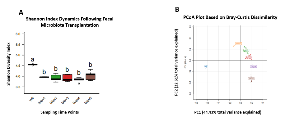
Figure A (Shannon index plot): Statistical comparisons were conducted via one-way ANOVA with Tukey’s multiple comparison post-hoc test, and the overall model P-value = 2 × 10⁻⁷. Groups labeled with distinct lowercase letters show significant intergroup differences at P < 0.05. W0 denotes the original human fecal microbiota inoculum before transplantation; BAW1–BAW5 represent mouse fecal microbiota collected at Weeks 1–5 post-fecal microbiota transplantation (FMT). The Shannon index of the W0 group was significantly higher than all post-transplant groups (P < 0.05), indicating host-selective colonization of intestinal microbes in mice due to interspecies incompatibility, leading to an overall reduction in gut microbial diversity after human FMT.
Figure B (PCoA plot): Principal Coordinate Analysis based on Bray-Curtis distance illustrating gut microbial community structure. PC1 and PC2 explain 44.43% and 22.61% of total microbial variance, respectively. Original W0 microbiota samples were completely separated from all post-transplant groups along the PC1 axis; BAW1 to BAW5 samples exhibited a sequential directional shift along the PC2 axis with prolonged housing time, demonstrating continuous ordered remodeling and gradual stabilization of mouse intestinal microbiota following FMT.

Anti-PD-1 Efficacy in GF vs. SPF Mice
Pharmacodynamic Evaluation of Anti-PD1 Antibody in Germ-Free vs SPF Tumor-Bearing Mice
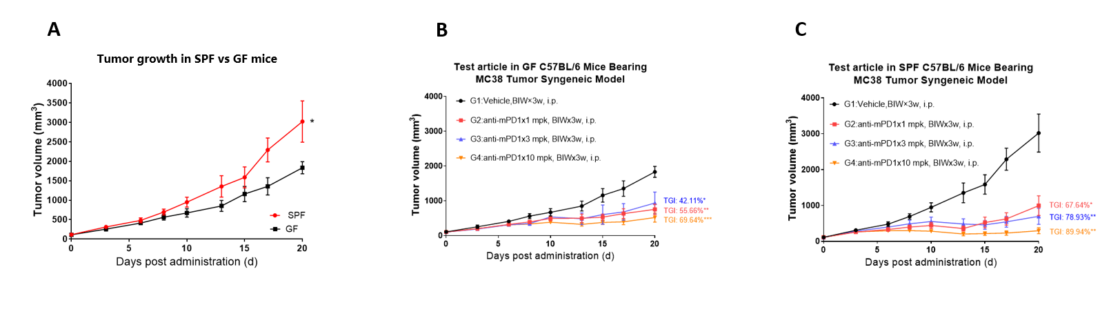
Figure A: Tumor growth curves of MC38 syngeneic tumors in SPF and germ-free (GF) C57BL/6 mice. Without drug intervention, SPF mice exhibited markedly faster tumor proliferation than GF mice, confirming that gut microbiota facilitates in vivo expansion of MC38 tumors.
Figure B: Tumor growth curves of GF tumor-bearing mice treated with gradient doses of anti-mPD1 antibody. G1 = vehicle control; G2/G3/G4 = 1, 3, 10 mg/kg anti-mPD1 administered intraperitoneally twice weekly for 3 consecutive weeks. Tumor growth inhibition (TGI) rose progressively with increasing dosage, demonstrating dose-dependent anti-tumor efficacy of anti-PD1 in GF mice.
Figure C: Tumor growth curves of SPF tumor-bearing mice treated with gradient anti-mPD1 doses. Grouping and administration regimens consistent with Figure B. Tumor growth inhibition rates across all dose groups were substantially higher than corresponding GF groups, revealing that gut microbiota markedly potentiates the anti-tumor activity of anti-PD1 immune checkpoint inhibitors.
Pharmacodynamic Evaluation of Colitis Treatment
Pharmacodynamic Assessment of 6-Thioguanine (6TG) and Akkermansia muciniphila in DSS-Induced Colitis in GF Mice
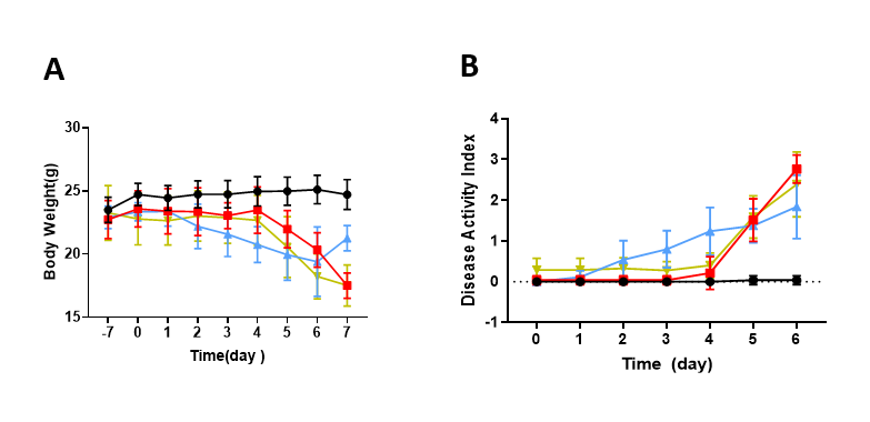
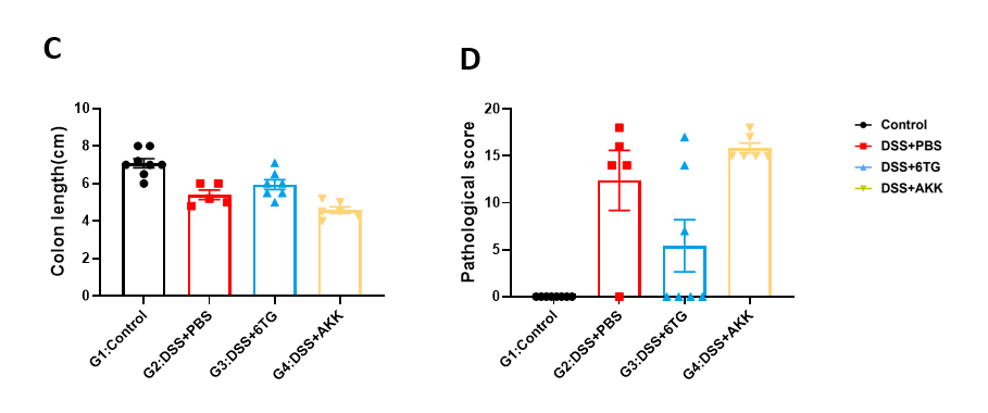
Figure A: Body weight dynamics of mice throughout the experimental period. G1 = untreated control; G2 = DSS-induced ulcerative colitis model group; G3 = positive drug intervention group (6TG); G4 = Akkermansia muciniphila intervention group. DSS administration induced significant weight loss in model mice, which was partially alleviated by both 6TG and Akkermansia treatment.
Figure B: Dynamic Disease Activity Index (DAI) scores across groups. DAI scores were significantly elevated in G2 relative to G1; treatment with 6TG and Akkermansia effectively alleviated colitis manifestations and reduced DAI values.
Figure C: Statistical quantification of colon length excised from sacrificed mice. Intergroup comparisons were performed via one-way ANOVA followed by Tukey’s post-hoc multiple comparison test, with statistical significance defined as P < 0.05. DSS exposure triggered colon shortening, while 6TG and Akkermansia supplementation attenuated colon atrophy.
Figure D: Histopathological injury scoring of colon tissues. One-way ANOVA with Tukey’s post-hoc test was used for statistical analysis, and differences with P < 0.05 were considered statistically significant. G2 displayed markedly elevated pathological scores compared with the healthy control group; both 6TG and Akkermansia intervention relieved colon tissue inflammatory damage.
All Product Lines



Ready to Accelerate Your Research?
Download



Newsletter Signup


We value your privacy
This website will store cookies on your computer. These cookies are used to collect information about how you interact with this website and enable us to remember you. We use this information to improve and customize your browsing experience, as well as to analyze and collect relevant data and metrics about visitors on this website and other platforms. For more information about the cookies we use, please refer to our privacy policy
If you decline, your information will not be tracked when visiting this website. A cookie will be used in your browser to remember your preference settings that you have not accepted.
Contact Us
Schedule a Meeting
Let us know which event you're attending and we'd be happy to connect with you.