


- By Indication
 Inflammatory and Autoimmune DiseasesRespiratory DiseasesInfectious DiseasesRare DiseaseGut Microbiota Research ServiceBy Modality
 Preclinical Model Strategies for T-Cell Engagers: From Oncology to Autoimmune Disease
 2026-10-27
 11:00 AM ET
 Dr. Wanjun Yuan & Dr. Haiqiao SunOne TCE Modality. Two Disease Contexts. Different Preclinical Questions.
- Animal Models
 KO/cKO Mouse


'/%3e%3c/svg%3e)
Acute Kidney Injury (AKI)
Why Choose Our AKI Models?
Integrated capabilities for acute kidney injury modeling and evaluation

Complementary Induction Approaches
Chemical induction with cisplatin and surgical induction by renal ischemia-reperfusion cover acute tubular injury, renal dysfunction, inflammation, and fibrotic remodeling.
01
Integrated Functional and Pathological Readouts
Functional endpoints — body weight, water intake, survival, serum creatinine, and BUN — are paired with kidney and lung histopathology.
02
Integrated Capabilities for Acute Kidney Injury Modeling and Evaluation
Our AKI platform combines cisplatin and renal ischemia–reperfusion models with BUN, creatinine, and histopathological assessments. Cisplatin studies characterize dose and time effects at 24–72 hours, while ischemia–reperfusion studies assess outcomes at Day 56. Semiquantitative tissue evaluation of KIM-1, F4/80, and COL3A1 provides complementary information on tubular injury, macrophage-associated inflammation, and collagen deposition, supporting model selection and endpoint planning for preclinical studies.
03
Model Library
Comprehensive AKI models for preclinical research

Case Studies & Validation Data
Cisplatin-Induced Acute Kidney Injury
Dose- and time-dependent kidney injury after a single cisplatin dose.

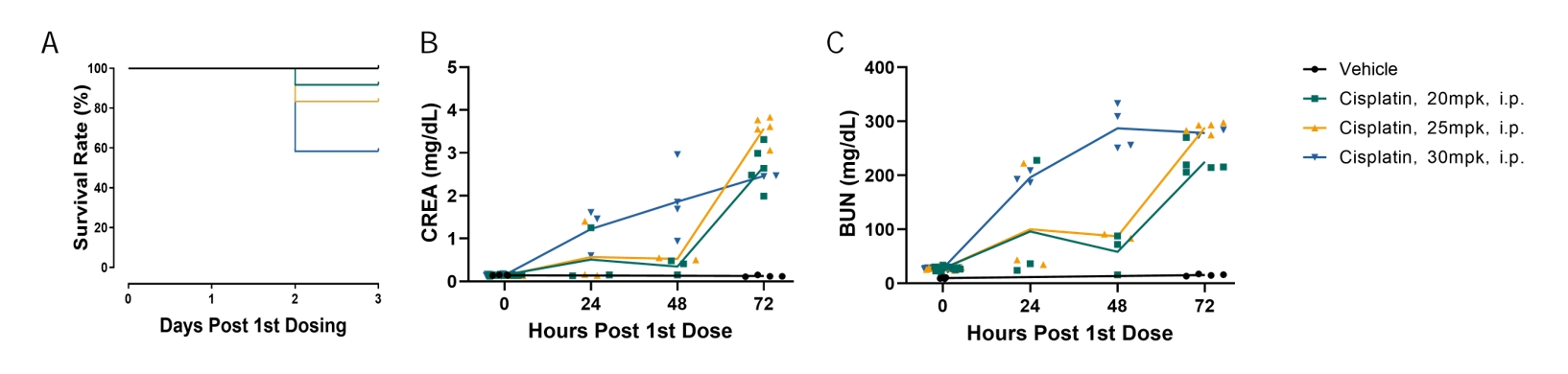
Cisplatin induced dose-dependent acute kidney injury and systemic toxicity, as demonstrated by reduced survival and rapid increases in serum creatinine and BUN. The 30 mg/kg group showed the greatest mortality and an earlier deterioration in renal function.
Figure 1. Dose-dependent induction of acute kidney injury by cisplatin in mice.
(A) Survival curves following a single intraperitoneal administration of vehicle or cisplatin at 20, 25, or 30 mg/kg. Survival decreased with increasing cisplatin dose, with the lowest survival observed in the 30 mg/kg group. (B) Serum creatinine (CREA) concentrations at 0, 24, 48, and 72 h after the first dose. (C) Blood urea nitrogen (BUN) concentrations at the indicated time points. Cisplatin treatment caused marked increases in serum CREA and BUN, indicating progressive renal dysfunction. Individual symbols represent individual animals, and lines connect group means. Group size: n = 4 for the vehicle group and n = 12 for each cisplatin group at study initiation.
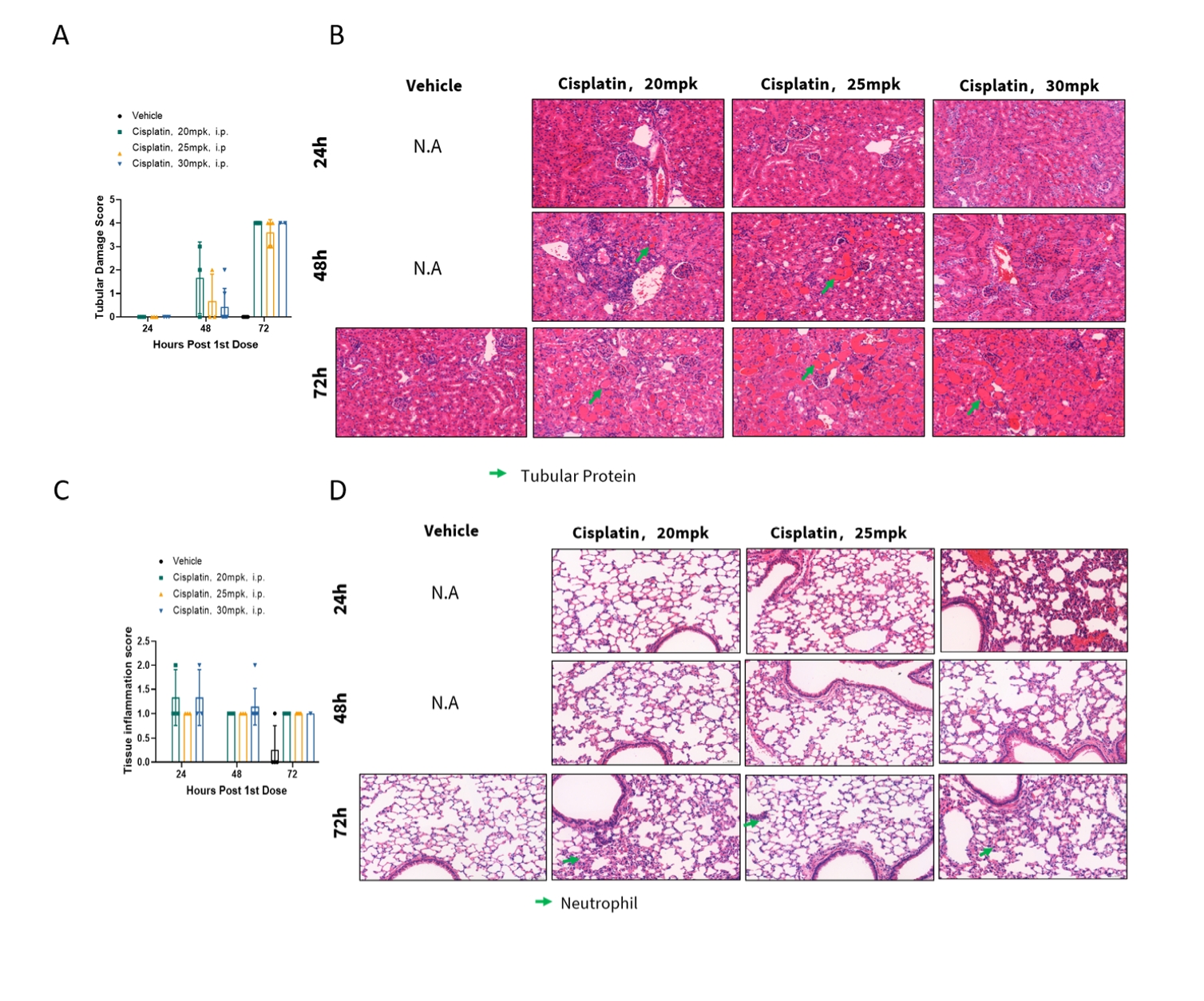
Cisplatin administration induced time-dependent renal tubular injury and pulmonary inflammation. Renal tubular damage became evident at 48 h and was severe by 72 h, accompanied by tubular protein accumulation. Inflammatory cell infiltration, including neutrophil accumulation, was also observed in the lung.
Figure 2. Histopathological evaluation of renal and pulmonary injury following cisplatin administration.
(A) Semiquantitative renal tubular damage scores at 24, 48, and 72 h after a single intraperitoneal administration of vehicle or cisplatin at 20, 25, or 30 mg/kg. (B) Representative H&E-stained kidney sections at the indicated time points. Cisplatin-treated mice exhibited progressive tubular injury and tubular protein accumulation, indicated by green arrows. (C) Semiquantitative lung tissue inflammation scores at 24, 48, and 72 h after treatment. (D) Representative H&E-stained lung sections showing inflammatory cell infiltration and neutrophil accumulation, indicated by green arrows. Individual symbols represent individual animals, and bars are presented as mean ± SEM. N.A., not available.

Ischemia-Reperfusion-Induced Kidney Injury
Long-term renal dysfunction, inflammation, and fibrosis following unilateral kidney ischemia-reperfusion injury
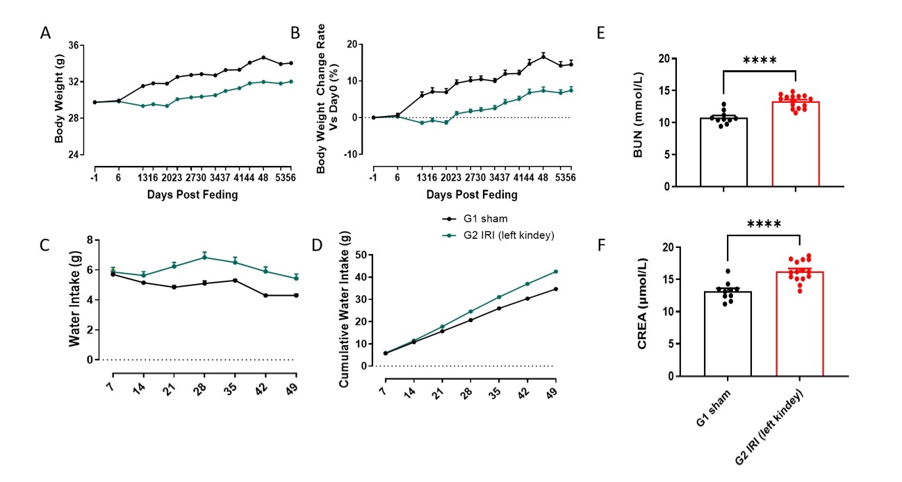
Unilateral left renal ischemia–reperfusion injury caused sustained physiological and renal functional abnormalities in mice. Compared with sham-operated mice, IRI mice showed reduced body weight gain, increased daily and cumulative water intake, and significantly elevated terminal serum BUN and creatinine levels.
Figure 1. Physiological and renal functional changes following unilateral renal ischemia–reperfusion injury in mice.
(A) Longitudinal changes in body weight; (B) body weight change rate relative to Day 0; (C) daily water intake; (D) cumulative water intake; (E) terminal serum blood urea nitrogen (BUN) levels at Day 56; and (F) terminal serum creatinine (CREA) levels at Day 56. Groups were defined as G1 Sham and G2 IRI (left kidney). Data presented as mean ± SD (n = 10–15 per group). Individual symbols in panels E and F represent individual animals. ****p < 0.0001.
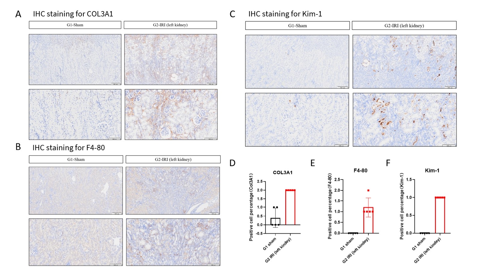
At Day 56, unilateral renal ischemia–reperfusion injury induced persistent renal fibrosis, macrophage infiltration, and tubular injury, as demonstrated by increased renal COL3A1, F4/80, and KIM-1 immunostaining compared with sham-operated mice.
Figure 2. Renal fibrosis, inflammation, and tubular injury following unilateral renal ischemia–reperfusion injury.
(A) Representative immunohistochemical staining for COL3A1 in kidney sections from G1 Sham and G2 IRI mice; (B) representative F4/80 immunohistochemical staining; (C) representative KIM-1 immunohistochemical staining; (D–F) semiquantitative scores for COL3A1, F4/80, and KIM-1 staining, respectively. Brown staining indicates positive immunoreactivity. Compared with the sham group, IRI mice showed increased COL3A1 deposition, F4/80-positive macrophage infiltration, and KIM-1 expression, indicating fibrotic remodeling, renal inflammation, and tubular injury. Scale bars: 200 μm in the upper-row images and 100 μm in the lower-row images. Data are presented as mean ± SD (n = 5 per group). Histological staining was graded according to the percentage of positive cells: 0, <5%; 1, 5%–<25%; 2, 25%–<50%; 3, 50%–<75%; and 4, ≥75%.
Product Lines


- 1
- 2
- 3
- 4
- 5
- 6
- 12
Recommended for You
Discover related models and services to accelerate your research

Accelerate Your Research
Download



Newsletter Signup


We value your privacy
This website will store cookies on your computer. These cookies are used to collect information about how you interact with this website and enable us to remember you. We use this information to improve and customize your browsing experience, as well as to analyze and collect relevant data and metrics about visitors on this website and other platforms. For more information about the cookies we use, please refer to our privacy policy
If you decline, your information will not be tracked when visiting this website. A cookie will be used in your browser to remember your preference settings that you have not accepted.
Contact Us
Schedule a Meeting
Let us know which event you're attending and we'd be happy to connect with you.