


- By Indication
 Inflammatory and Autoimmune DiseasesRespiratory DiseasesInfectious DiseasesRare DiseaseGut Microbiota Research ServiceBy Modality
 Preclinical Model Strategies for T-Cell Engagers: From Oncology to Autoimmune Disease
 2026-10-27
 11:00 AM ET
 Dr. Wanjun Yuan & Dr. Haiqiao SunOne TCE Modality. Two Disease Contexts. Different Preclinical Questions.
'/%3e%3c/svg%3e)

Sarcopenia
- 4Sarcopenia Model Options
- 6Key Skeletal Muscle Groups
- 10+Validated Functional & Compositional Indicators
Our Advantages

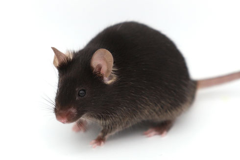
Diverse and Clinically-Oriented Sarcopenia Models
Our sarcopenia models cover dexamethasone-induced, hindlimb suspension, orchiectomy, and combined orchiectomy-hindlimb suspension models, designed to mimic varied clinical pathogenic mechanisms of sarcopenia for translational research development.
01
Comprehensive Phenotypic & Functional Assessment
Multi-parameter evaluation including qNMR body composition analysis, DEXA scanning, grip strength measurement, muscle endurance testing, behavioral assessment, biomarker detection, and histopathological examination.
02
Flexible and Customizable Study Design
Adaptable study designs tailored to diverse sarcopenia-related research and drug development needs, with customizable induction protocols and standardized efficacy evaluation workflows to deliver high-quality testing
03
Model Library

Case Studies & Validation Data
Explore our validated sarcopenia studies demonstrating the efficacy of various therapeutic interventions.
Dexamethasone induced Sarcopenia Model
Efficacy test of Bimagrumab on Dexamethasone induced sarcopenia model.

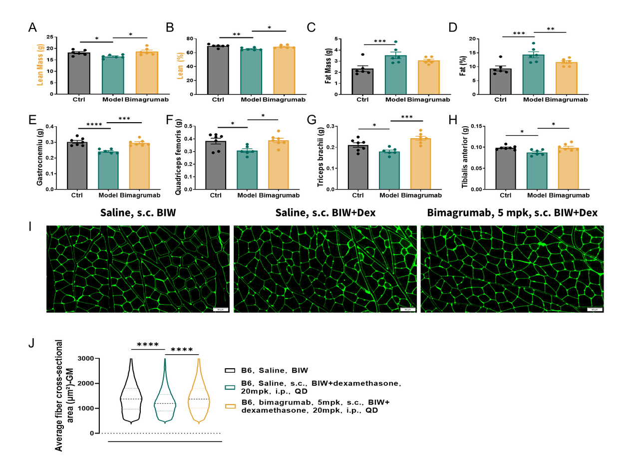 These results indicate that Bimagrumab improves selected body composition, muscle-related parameters, and muscle fiber morphology in the dexamethasone-induced sarcopenia model, supporting its utility for pharmacological evaluation in sarcopenia and muscle wasting studies.
(A–D) Quantitative analysis of body composition and muscle-related parameters, including lean mass, lean mass percentage, and other skeletal muscle phenotype-associated indicators. Compared with the dexamethasone model group, bimagrumab treatment showed improving trends in selected body composition and muscle-related parameters, suggesting a potential protective effect against dexamethasone-induced muscle loss. (E–H) Quantitative analysis of skeletal muscle. Bimagrumab treatment partially improved muscle phenotype-related readouts compared with the model group, indicating its potential to alleviate dexamethasone-induced skeletal muscle atrophy. (I) Representative fluorescence images of skeletal muscle cross-sections. Green fluorescence outlines the muscle fiber borders and was used to assess changes in muscle fiber morphology and size among groups. Scale bar: 50 μm. (J) Distribution analysis of muscle fiber cross-sectional area. Compared with the examethasone model group, bimagrumab treatment showed an improving trend in muscle fiber size distribution, suggesting partial attenuation of dexamethasone-induced muscle fiber atrophy. Data are presented as mean ± SEM.
Hindlimb Suspension-Induced Sarcopenia Model
For this model, mice are anesthetized supine. Bilateral hind limbs are wrapped with non-elastic bandages in plantar flexion, stabilized by vinyl-coated steel wire. Softened bandages and triangular gypsum supports are applied for fixation and necrosis monitoring.
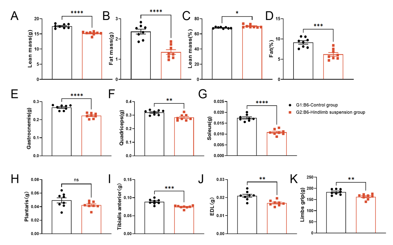
These results indicate that hindlimb suspension induces significant alterations in body composition, skeletal muscle mass, and muscle function‑related parameters in C57BL/6 mice, supporting the reliability of this model for sarcopenia and disuse‑induced muscle wasting research.
(A-D) Quantitative analysis of body composition parameters, including lean mass, fat mass, lean mass percentage, and fat percentage. Compared with the control group, the hindlimb suspension group exhibited markedly decreased lean mass and lean mass percentage, alongside reduced fat mass and fat percentage, reflecting disrupted whole‑body body composition induced by mechanical unloading. (E-J) Quantitative analysis of individual skeletal muscle weight. Hindlimb suspension significantly reduced the weight of gastrocnemius, quadriceps, soleus, tibialis anterior, and extensor digitorum longus (EDL) muscles, while no significant difference was observed in plantaris muscle weight, indicating widespread skeletal muscle atrophy induced by hindlimb unloading. (K) Assessment of limb grip strength. Compared with the control group, the hindlimb suspension group displayed significantly decreased limb grip strength, demonstrating impaired muscle function under disuse conditions. Data are presented as mean ± SEM. *p<0.05, **p<0.01, ***p<0.001, ****p<0.0001; ns, not significant.
Orchiectomy-induced Sarcopenia Model
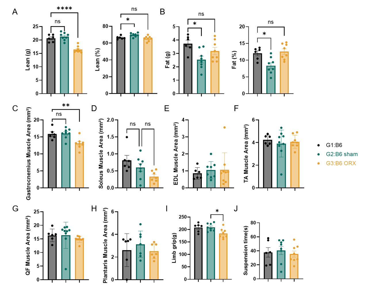 These results indicate that orchiectomy induces characteristic changes in body composition, skeletal muscle morphology, and muscle function in C57BL/6 mice, verifying the validity of this orchiectomy-induced sarcopenia model for preclinical efficacy evaluation.
(A-B) Quantitative analysis of body composition parameters including lean mass, lean percentage, fat mass and fat percentage. Compared with the sham-operated group, the orchiectomy (ORX) group showed significantly reduced lean mass, accompanied by altered fat percentage, reflecting systemic metabolic remodeling triggered by androgen deficiency. (C-H) Quantitative analysis of crosssectional areas of multiple skeletal muscles. ORX treatment significantly decreased crosssectional areas of the soleus muscle, while no statistically significant differences were observed in the tibialis anterior (TA), extensor digitorum longus (EDL), quadriceps femoris (QF), and plantaris muscles, indicating musclespecific atrophy induced by androgen deprivation. (I-J) Functional assessment of skeletal muscle. Compared with the sham group, the ORX group exhibited significantly reduced limb grip strength and a decreasing trend in suspension time, demonstrating impaired muscle function under orchiectomy conditions. Data are presented as mean ± SEM. *p<0.05, **p<0.01, ****p<0.0001; ns, not significant.

All Product Lines


- 1
- 2
- 3
- 4
- 5
- 6
- 12
Recommended for You
Discover related models and services to accelerate your research

Accelerate Your Research
Download



Newsletter Signup


We value your privacy
This website will store cookies on your computer. These cookies are used to collect information about how you interact with this website and enable us to remember you. We use this information to improve and customize your browsing experience, as well as to analyze and collect relevant data and metrics about visitors on this website and other platforms. For more information about the cookies we use, please refer to our privacy policy
If you decline, your information will not be tracked when visiting this website. A cookie will be used in your browser to remember your preference settings that you have not accepted.


Contact Us
Schedule a Meeting
Let us know which event you're attending and we'd be happy to connect with you.