


'/%3e%3c/svg%3e)
- By Indication
 Inflammatory and Autoimmune DiseasesRespiratory DiseasesInfectious DiseasesRare DiseaseGut Microbiota Research ServiceBy Modality
 From Bench to Bedside: Modeling Autoimmune, Genetic, and Fibrotic Kidney Diseases in Mice
 2026-09-22
 11:00 AM ET
 Dr. Yinlian ZhangStudy Director, Autoimmune Pipeline

'/%3e%3c/svg%3e)
'/%3e%3c/svg%3e)
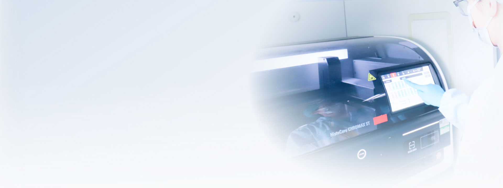
Preclinical Imaging and Analysis Services

Dual-Energy X-ray Absorptiometry (DEXA)
High-resolution Bone Density Assessment
High-resolution X-ray scanning enables whole-body and regional assessment of bone mineral density and changes in bone structure, supporting efficacy evaluation in osteoporosis and other bone metabolism studies.
Body Composition Analysis
Quantitative analysis of fat mass, lean tissue, and body composition helps distinguish the sources of weight change and evaluate treatment effects in metabolic disease studies.
In Vivo and Ex Vivo Imaging
DEXA supports both in vivo animal imaging and ex vivo tissue analysis, enabling researchers to correlate longitudinal phenotypic changes with tissue-level findings.
Longitudinal Studies
Noninvasive imaging enables repeated measurements in the same animals over time, supporting longitudinal analysis while potentially reducing the number of animals required.
In Vivo Optical Imaging
The in vivo optical imaging system captures and quantifies bioluminescent and fluorescent signals in living organisms, enabling real-time, non-invasive monitoring of tumor progression, metastatic spread, drug distribution, and various physiological processes in mouse models. The platform supports drug efficacy evaluation and pharmacokinetic analysis.
Ultrasound Imaging and Analysis
Cardiac Ultrasound Imaging
Cardiac Structure Assessment: High-resolution ultrasound imaging measures parameters including left ventricular internal diameter and ventricular wall thickness to assess cardiac size and structure.
Cardiac Function Assessment: Measurement of parameters including left ventricular ejection fraction and fractional shortening supports evaluation of systolic and diastolic cardiac function.
Organ Ultrasound Imaging
Liver Ultrasound: Assessment of liver size, shape, and internal echogenicity supports the detection and evaluation of liver abnormalities.
Kidney Ultrasound: Assessment of kidney size, cortical thickness, and structural abnormalities such as hydronephrosis supports evaluation of renal structure.
Blood Flow Analysis
Carotid Artery Blood Flow Detection: Coronary artery blood flow measurements support cardiovascular disease studies and evaluation of therapeutic effects.
Coronary Artery Blood Flow Detection: Echocardiography enables measurement of coronary artery blood flow to support cardiovascular disease research and evaluation of therapeutic effects.
Noninvasive Blood Pressure Measurement
Tail Artery Blood Pressure Measurement: Noninvasive tail artery measurement enables longitudinal monitoring of blood pressure in mice, including changes associated with drug intervention and disease progression.
Ultrasound Data Analysis
The ultrasound analysis platform provides high-precision image recognition and quantitative data analysis. Measurements including organ dimensions and hemodynamic parameters support detailed assessment of cardiovascular function and organ status in mouse models.
- Dual-Energy X-ray Absorptiometry (DEXA) Platform
① Platform Advantages
Precision: High-resolution measurement of bone mineral density and body composition
Noninvasive Analysis: Minimal animal handling for repeated measurements
Reproducibility: Consistent data across longitudinal studies
Image Quality: High-resolution imaging for detailed anatomical and body composition analysis
② Imaging Capabilities
The system generates comprehensive imaging outputs, including high-resolution X-ray images, bone mineral density (BMD) maps, and pseudo-colored body composition visualizations enabling multi-dimensional analysis in a single scan.
X-Ray Imaging: High-resolution radiographs support precise measurement of regional bone length, fracture assessment, and implant localization with exceptional clarity.
Bone Mineral Density Mapping: Quantitative BMD visualization highlights density distribution across skeletal regions, facilitating early detection of bone loss and treatment response evaluation.
Body Composition Analysis: Pseudo-colored imaging distinguishes fat, lean tissue, and bone mass, providing an intuitive overview of physiological changes in metabolic and aging studies.


X-ray images BMD Images BMD Images BMD
BMC
Bone Area
Tissue Area
Fat(%%)
Fat(g)
Lean(g)
0.087
0.553
6.33
16.507
14.104
3.294
20.061


Isolated bone tissue
BMD
BMC
Bone Area
Tissue Area
Fat(%)
Fat(g)
Lean(g)
0.034
0.001
0.017
0.267
4
0.005
0.128
0.016
0.004
0.22
3.181
32.037
0.591
1.253
- <i>In Vivo</i> Optical Imaging Platform
① Platform Advantages
Broad Coverage: Large field of view for simultaneous imaging of multiple regions
High Throughput: Multi-animal imaging for efficient study execution
Multimodal Imaging: Integrated fluorescence and bioluminescence capabilities
Quantitative Analysis: Absolute signal intensity quantification for rigorous data analysis
② Imaging and Analysis Capabilities
In vivo optical imaging enabled real-time monitoring of drug distribution, accumulation, and clearance in live animals across multiple time points.
- Ultrasound and Cardiovascular Imaging Platform
① Noninvasive Blood Pressure Measurement: Kent CODA system
② Small-Animal Ultrasound Imaging: VINNO VINNOD8 LAB system

③ Animal Electrophysiology: ADInstruments system


④ Laser Speckle Blood Flow Imaging: RWD RFLSI system


Ready to Accelerate Your Research?


'%3e%3cpath%20d='M13.6882%2012L23.6426%202.06059C23.8678%201.83321%2023.9941%201.52611%2023.9941%201.20607C23.9941%200.886035%2023.8678%200.578937%2023.6426%200.351553C23.4152%200.126341%2023.1081%200%2022.7881%200C22.468%200%2022.1609%200.126341%2021.9335%200.351553L11.9941%2010.306L2.05473%200.351553C1.82735%200.126341%201.52025%200%201.20021%200C0.880175%200%200.573077%200.126341%200.345693%200.351553C0.120482%200.578937%20-0.00585938%200.886035%20-0.00585938%201.20607C-0.00585937%201.52611%200.120482%201.83321%200.345693%202.06059L10.3001%2012L0.345693%2021.9394C0.120482%2022.1668%20-0.00585937%2022.4739%20-0.00585938%2022.7939C-0.00585938%2023.114%200.120482%2023.4211%200.345693%2023.6484C0.573077%2023.8737%200.880175%2024%201.20021%2024C1.52025%2024%201.82735%2023.8737%202.05473%2023.6484L11.9941%2013.694L21.9335%2023.6484C22.1666%2023.8397%2022.4624%2023.9374%2022.7635%2023.9226C23.0646%2023.9079%2023.3494%2023.7816%2023.5626%2023.5684C23.7757%2023.3553%2023.902%2023.0704%2023.9168%2022.7694C23.9316%2022.4683%2023.8338%2022.1724%2023.6426%2021.9394L13.6882%2012Z'%20fill='black'%20fill-opacity='0.96'/%3e%3c/g%3e%3cdefs%3e%3cclipPath%20id='clip0_2458_12695'%3e%3crect%20width='24'%20height='24'%20fill='white'/%3e%3c/clipPath%3e%3c/defs%3e%3c/svg%3e)
Submit Inquiry
Contact Us
Schedule a Meeting
Let us know which event you're attending and we'd be happy to connect with you.
Download
Download
Watch On-Demand Webinar
Newsletter Signup
'%3e%3cpath%20d='M13.6882%2012L23.6426%202.06059C23.8678%201.83321%2023.9941%201.52611%2023.9941%201.20607C23.9941%200.886035%2023.8678%200.578937%2023.6426%200.351553C23.4152%200.126341%2023.1081%200%2022.7881%200C22.468%200%2022.1609%200.126341%2021.9335%200.351553L11.9941%2010.306L2.05473%200.351553C1.82735%200.126341%201.52025%200%201.20021%200C0.880175%200%200.573077%200.126341%200.345693%200.351553C0.120482%200.578937%20-0.00585938%200.886035%20-0.00585938%201.20607C-0.00585937%201.52611%200.120482%201.83321%200.345693%202.06059L10.3001%2012L0.345693%2021.9394C0.120482%2022.1668%20-0.00585937%2022.4739%20-0.00585938%2022.7939C-0.00585938%2023.114%200.120482%2023.4211%200.345693%2023.6484C0.573077%2023.8737%200.880175%2024%201.20021%2024C1.52025%2024%201.82735%2023.8737%202.05473%2023.6484L11.9941%2013.694L21.9335%2023.6484C22.1666%2023.8397%2022.4624%2023.9374%2022.7635%2023.9226C23.0646%2023.9079%2023.3494%2023.7816%2023.5626%2023.5684C23.7757%2023.3553%2023.902%2023.0704%2023.9168%2022.7694C23.9316%2022.4683%2023.8338%2022.1724%2023.6426%2021.9394L13.6882%2012Z'%20fill='%23008077'%20fill-opacity='0.96'/%3e%3c/g%3e%3cdefs%3e%3cclipPath%20id='clip0_2458_12695'%3e%3crect%20width='24'%20height='24'%20fill='white'/%3e%3c/clipPath%3e%3c/defs%3e%3c/svg%3e)
We value your privacy
This website will store cookies on your computer. These cookies are used to collect information about how you interact with this website and enable us to remember you. We use this information to improve and customize your browsing experience, as well as to analyze and collect relevant data and metrics about visitors on this website and other platforms. For more information about the cookies we use, please refer to our privacy policy
If you decline, your information will not be tracked when visiting this website. A cookie will be used in your browser to remember your preference settings that you have not accepted.

