


'/%3e%3c/svg%3e)

- By Indication
 Inflammatory and Autoimmune DiseasesRespiratory DiseasesInfectious DiseasesRare DiseaseGut Microbiota Research ServiceBy Modality
 Preclinical Model Strategies for T-Cell Engagers: From Oncology to Autoimmune Disease
 2026-10-27
 11:00 AM ET
 Dr. Wanjun Yuan & Dr. Haiqiao SunOne TCE Modality. Two Disease Contexts. Different Preclinical Questions.

'/%3e%3c/svg%3e)
'/%3e%3c/svg%3e)

Autoimmune
Systemic Lupus Erythematosus (SLE): Pathogenesis, Animal Models & Preclinical Drug Selection
Systemic lupus erythematosus (SLE) is a heterogeneous autoimmune rheumatic disease. The core pathology is the breakdown of immune self‑tolerance: autoreactive B cells produce autoantibodies, immune complexes deposit in multiple organs, and innate immune pathways such as the cGAS‑STING and TLR7‑type I IFN axis are persistently activated. Human SLE has highly variable clinical manifestations, which create major challenges for drug development. GemPharmatech has established a comprehensive panel of SLE models covering diverse pathogenic mechanisms, to support evaluation of different drug modalities and targets. Read the whole blog to learn more.
 2026-10-21

Systemic lupus erythematosus (SLE) is a heterogeneous autoimmune rheumatic disease. The core pathology is the breakdown of immune self‑tolerance: autoreactive B cells produce autoantibodies, immune complexes deposit in multiple organs, and innate immune pathways such as the cGAS‑STING and TLR7‑type I IFN axis are persistently activated.
Human SLE has highly variable clinical manifestations, which create major challenges for drug development. No single animal model can fully recapitulate all human disease features. Instead, each model amplifies one specific pathogenic axis, making model selection based on your drug’s mechanism of action the critical step for reliable preclinical assessment. GemPharmatech has established a comprehensive panel of SLE models covering diverse pathogenic mechanisms, to support evaluation of different drug modalities and targets. Below is a list of SLE models developed by GemPharmatech, along with their pathogenic mechanisms and applications in drug assessment.
Model | Mechanism | Disease Phenotype | Application |
B6-hBAFF (APRIL,CD3, BCMA,CD20,CD19) | Activated B cells | Increased B cell population, increased antibodies, proteinuria, lupus nephritis | Fusion protein: Telitacicept Small molecules: BTK inhibitors T Cell Engagers: Blincyto, Teclistamab, Mosunetuzumab |
B6-Trex1 KO (SLE/CLE) | cGAS-STING-IFNα | Increased anti-dsDNA IgG, ALT, ISD, tissue fibrosis | Small molecules: inhibitor for cGAS-STING pathways Antibodies: anti-IFNAR |
BALB/c-Tlr7-p.Y264H | TLR7 gain of function mutation | Increased anti-dsDNA IgG, nephritis, increased B cell population, ISD | Small molecules: Enpatoran (TLR7 inhibitor), BTK inhibitors |
TLR7 agonist-induced | TLR7 pathway activation | Increased B cell population, anti-dsDNA IgG, nephritis, proteinuria | Antibodies: B cell targeting antibodies Small molecules: Enpatoran |
SLE patient PBMC-NCG | Pathogenic B cells from patients | Increased anti-dsDNA IgG, B cell population, IgG deposition in kidney | T Cell Engagers: Teclistamab, Blincyto CAR-T: ex vivo, in vivo Antibodies: Rituximab, Ocrelizumab |
HSC reconstituted (HSC-NCG-M) | TLR7 induced immune cell activation | IgG deposition in kidney, antibodies | T Cell Engagers: Blincyto CAR-T: ex vivo, in vivo Antibodies: Rituximab |
Pristane induced | Activation of peritoneal macrophages | Increased autoantibodies, ISD | Antibody Small molecules: Enpatoran, Upadacitinib |
Breakdown of Above SLE Animal Models
B6‑hBAFF (APRIL, CD3, BCMA, CD20, CD19, APRIL)
Mechanism: Overactivated B cells via BAFF signaling
Disease phenotype: Elevated autoantibodies and B cell population, proteinuria, lupus nephritis
Application: Can be crossed with CD3, BCMA, CD19, CD20 humanized strains to introduce human target genes into this line, enabling evaluation of the efficacy of anti-human antibodies.
Benefits: Ideal for B‑cell‑targeted biologics. It recapitulates autoantibody production and kidney injury, the key clinical endpoint of lupus.
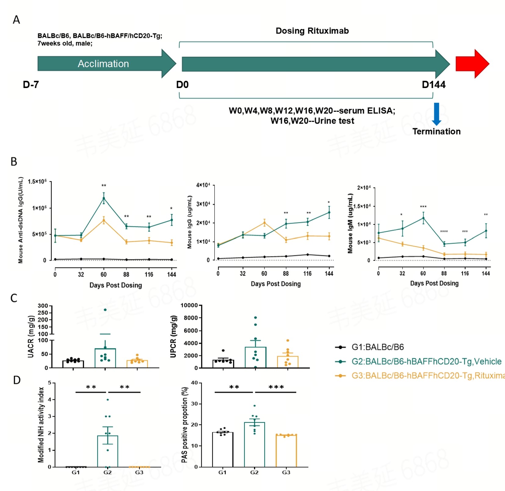
Study design for rituximab evaluation in the B6-hBAFF/hCD20 humanized mouse model of SLE(A). Rituximab treatment significantly reduced serum levels of anti-dsDNA IgG, total IgG, and IgM(B). Urinary albumin-to-creatinine ratio (UACR) and urinary protein-to-creatinine ratio (UPCR) displayed numerical decreases following rituximab treatment (C), but these decreases did not reach statistical significance. Rituximab treatment reduced both the lupus nephritis activity index and the proportion of PAS-positive staining(D). Statistical analysis was performed using one-way ANOVA, and all data are presented as mean ± SEM. *p<0.05, **p<0.01, ***p<0.001,****p<0.0001
B6‑Trex1 KO (SLE/CLE)
Mechanism: cGAS‑STING‑IFNα pathway activation. Trex1 is a DNA exonuclease; loss of function leads to accumulated intracellular self‑DNA.
Disease Phenotype: Anti‑dsDNA IgG, elevated ALT, ISD, tissue fibrosis
Application: Small molecule inhibitors or antibodies targeting cGAS/STING/IFN axis
Benefits: Perfect for innate immune small molecules focused on nucleic acid sensing and tissue fibrosis.
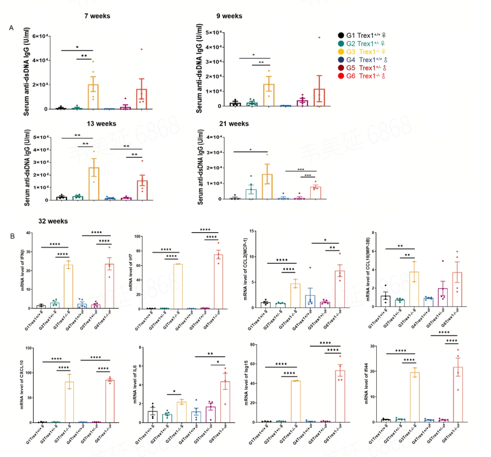
In both female and male mice aged 7 to 21 weeks, serum anti-dsDNA levels were significantly higher in B6-Trex1-KO mice compared to control B6 mice(A). This result aligns with the previously described autoimmune phenotype of TREX1-deficient mice, where genomic DNA accumulation triggers autoimmune responses characteristic of SLE pathogenesis. An increased interferon-stimulatory DNA (ISD) response was observed in heart tissue of TREX-1 KO mice at 32 weeks of age(B). Statistical analysis was performed using one-way ANOVA, and all data presented as mean ± SEM. N=5 per group*p<0.05, **p<0.01, ***p<0.001,****p<0.0001
Mechanism: Tlr7 gain-of-‑function mutation
Disease Phenotype: Anti‑dsDNA IgG, nephritis, altered B cell populations, ISD
Application: Small‑molecule TLR7 inhibitors or molecules targeting interferon.
Benefits: Genetically driven TLR7 hyperactivity, mimics the human SLE IFN signature. Great for mechanistic validation of TLR7‑targeted small molecules.
Mechanism: Pharmacological activation of the TLR7 pathway
Disease Phenotype: Increased anti‑dsDNA IgG and B cell population, nephritis, proteinuria
Application: Antibodies or small molecules against B cells, TLR7 (e.g. Enpatoran) or interferon.
Benefits: Inducible model with rapid disease onset. Suitable for screening TLR7 pathway inhibitors and agents that block downstream B cell activation.
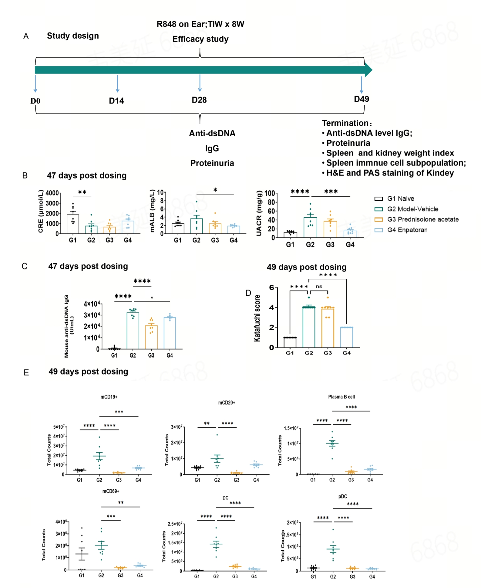
Study design of TLR7 agonist-induced SLE mouse model(A). Enpatoran reduced urinary protein levels at Day 47 post-dose(B). Anti-dsDNA IgG levels were decreased following 47 days of prednisolone treatment(C). Renal pathological scores were reduced at Day 49 post Enpatoran administration(D). Splenic B cells and DCs were decreased after 49 days of treatment with prednisolone and Enpatoran(E). Statistical analysis was performed using one‑way ANOVA, and all data presented as mean ± SEM. N=8 per group. *p<0.05, **p<0.01, ***p<0.001,****p<0.0001
Mechanism: Patient‑derived pathogenic B cells engrafted into immunodeficient NCG mice
Disease Phenotype: Anti‑dsDNA IgG, abnormal B cell populations, IgG deposition in kidney
Application: TCE (Teclistamab,Blincyto ), autologous/in vivo CAR‑T, anti‑CD20 antibodies (Rituximab, Ocrelizumab)
Benefits: Humanized model with patient pathogenic immune cells. Unique platform for human cell therapies, which do not cross‑react with murine B cells.
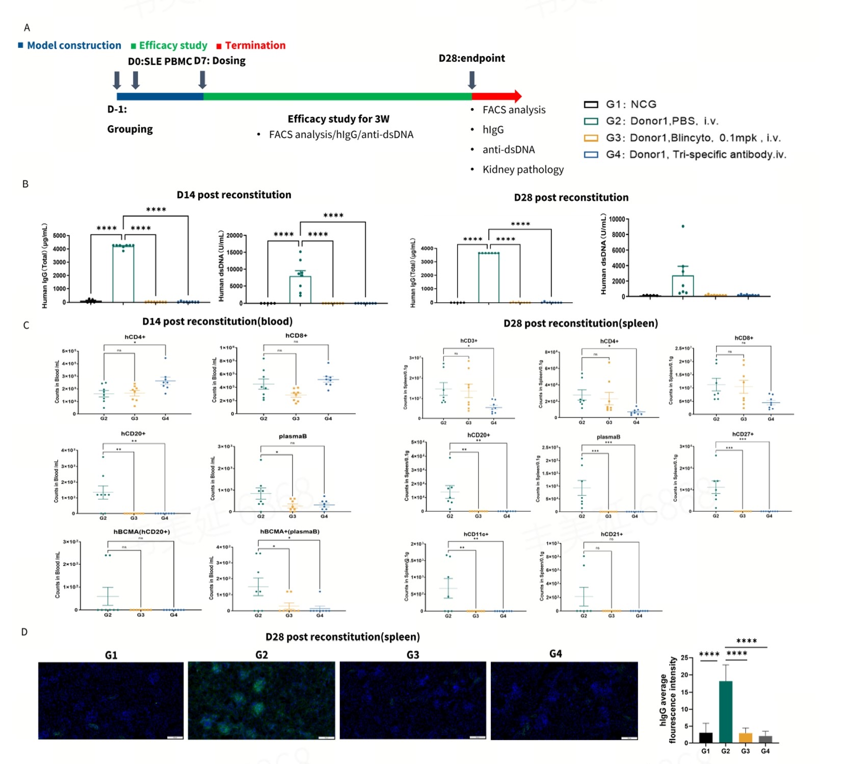
Study design of SLE patient PBMC‑NCG model (A). Blincyto and the trispecific TCE significantly reduced serum total IgG and anti-dsDNA IgG levels (B). Blincyto and the trispecific TCE test article significantly decreased B cell and plasma cell numbers (C). Blincyto and the test article markedly reduced IgG deposition in the kidney. Statistical analysis was performed using one‑way ANOVA, and all data presented as mean ± SEM. N=8 per group. *p<0.05, **p<0.01, ***p<0.001,****p<0.0001.
HSC reconstituted (HSC‑NCG‑M)
Mechanism: HSC reconstitution gives human immune cells with TLR7‑driven immune activation
Disease Phenotype: Human immune cell populations, kidney IgG deposition, autoantibodies
Application: Evaluation of B cell targeting therapy, e.g. TCE (Blincyto), CAR‑T, Rituximab etc.
Benefits: Multi lineage human immune system. Better suited for longer‑term study of human immune cell‑mediated disorders.
Pristane induced model
Mechanism: Activation of peritoneal macrophages
Disease Phenotype: Autoantibodies, ISD
Application: Antibodies and small molecules that target B cells and interferon
Benefits: Low cost, rapid induction. Good for preliminary compound screening, but only mild disease; not ideal for studying severe progressive nephritis.
Key caveat: Each model only represents one slice of SLE pathogenesis. Positive efficacy in one model does not guarantee clinical success. A tiered strategy combining multiple complementary models is recommended to de‑risk preclinical data.
Closing Thoughts
SLE is a multi‑pathway autoimmune disease. Preclinical teams need to select SLE models based on the molecular mechanism of their therapeutic candidate and the primary study endpoints: autoantibody levels, B cell counts, kidney IgG deposition, proteinuria, tissue fibrosis or ISD.
The table of SLE models provides a comprehensive review of various SLE models based on mechanism of disease pathogenesis and guidance for model selection depending on the mechanism of action of the therapeutic candidates. By aligning the model’s pathogenic axis with your drug target, you can generate more translatable data to support clinical development.
What is your SLE drug target? Which model are you planning to test first? Contact us today!

Share


Recommended for You
More infomation about blogs

Find Models


Submit Inquiry
Contact Us
Schedule a Meeting
Let us know which event you're attending and we'd be happy to connect with you.
Download
Download
Watch On-Demand Webinar
Newsletter Signup

We value your privacy
This website will store cookies on your computer. These cookies are used to collect information about how you interact with this website and enable us to remember you. We use this information to improve and customize your browsing experience, as well as to analyze and collect relevant data and metrics about visitors on this website and other platforms. For more information about the cookies we use, please refer to our privacy policy
If you decline, your information will not be tracked when visiting this website. A cookie will be used in your browser to remember your preference settings that you have not accepted.

