

Anti-CD40 Induced Colitis Models
Model strategy
Experimental mouse strains: RAG-/-, 6-7weeks old, female
Modeling reagents: anti-CD40 antibody
Modeling method:
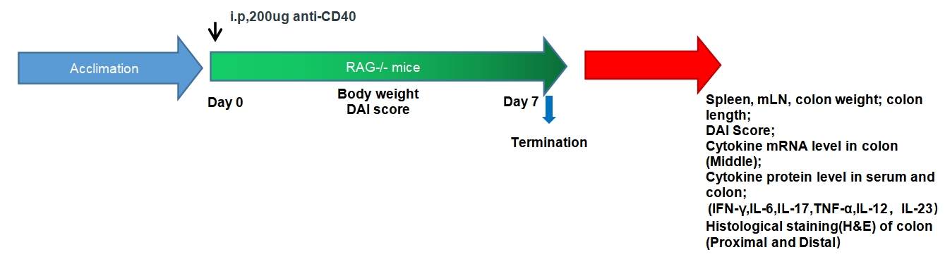
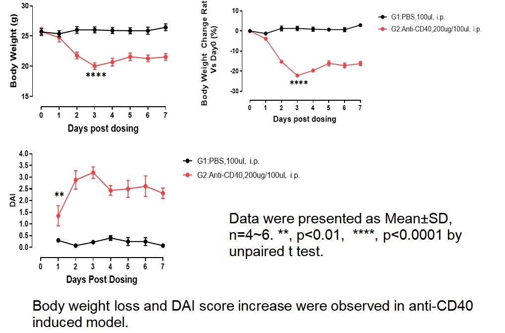
Short modeling time (7 days), ease of use (upon injection of anti-CD40 antibody);
Evaluate the innate immune response in disease using cytokine profiling of : IFN-γ、IL-12、IL-23、TNF-α.
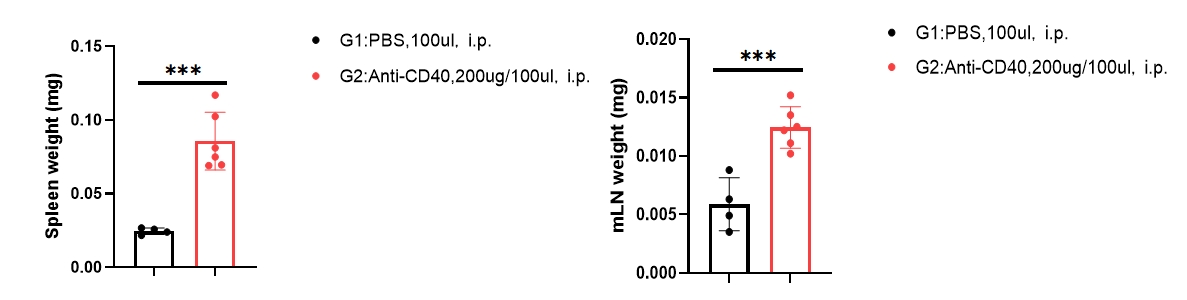
Spleen and mesenteric lymph node(mLN) weight increase was observed in anti-CD40 induced model.
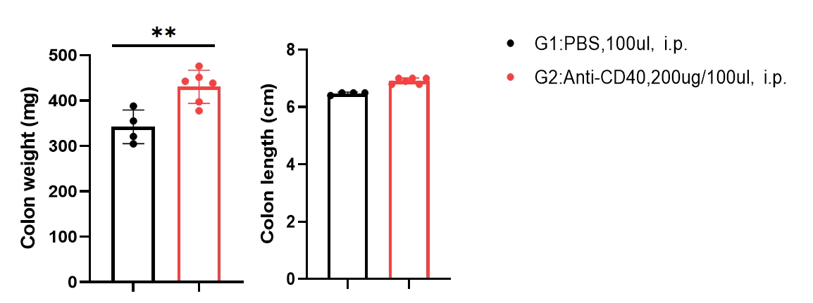
Spleen, mesenteric lymph node (mLN) and colon weight increase was observed in anti-CD40 induced model.
ELISA
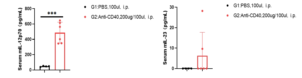
Cytometric bead array
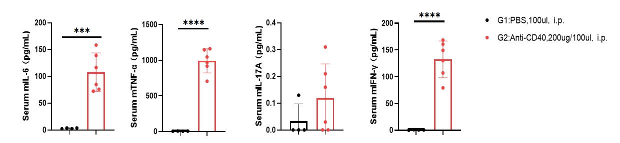
Serum IL-12, IL-23, IL-6,TFN-α and IFN-γ levels were elevated in anti-CD40-induced model.
Cytometric bead array
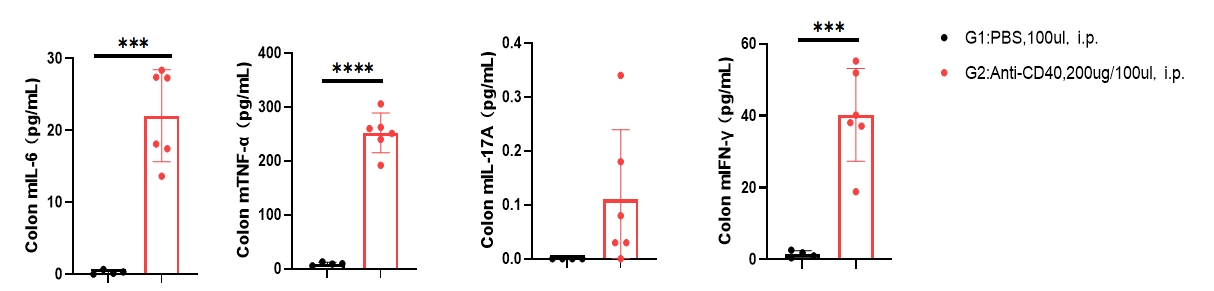
Colon IL-6,TFN-α and IFN-γ cytokines protein level was significantly increased in anti-CD40-induced model.
RT-QPCR
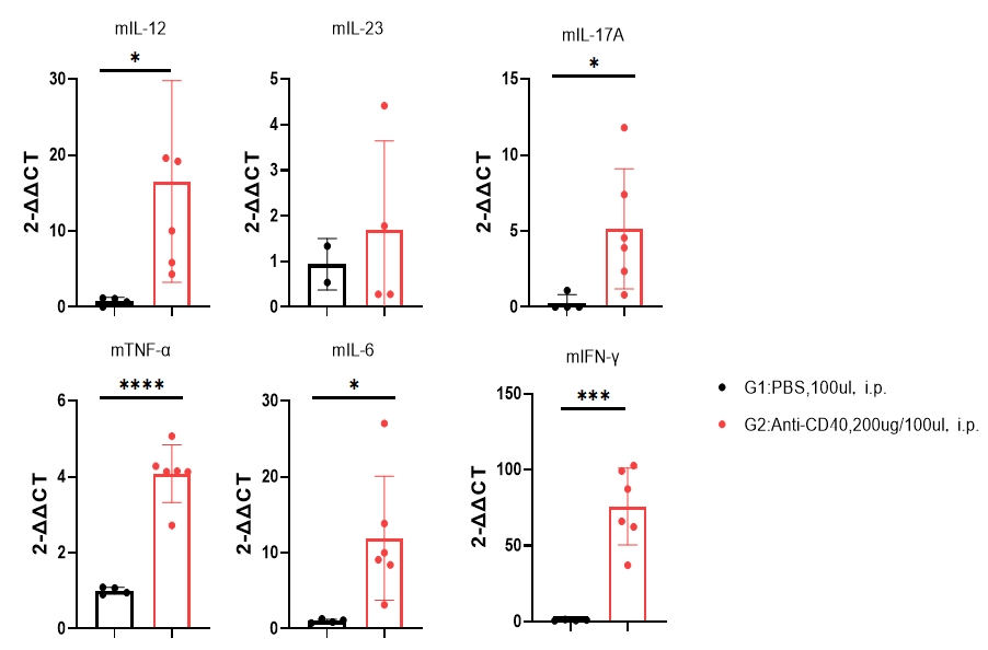
Colon IL-12, IL-6,TFN-α, IL17A and IFN-γ cytokines mRNA level was significantly increased in anti-CD40 induced model.
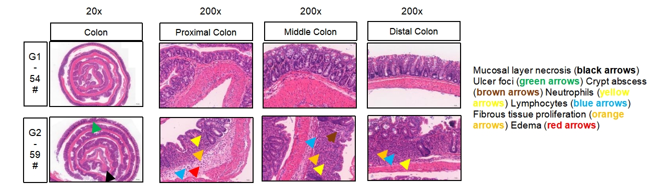
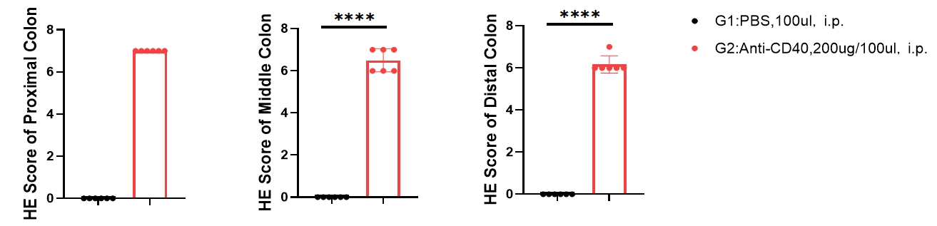
H&E staining of colon demonstrating histopathological features associated with anti-CD40 induced colitis