

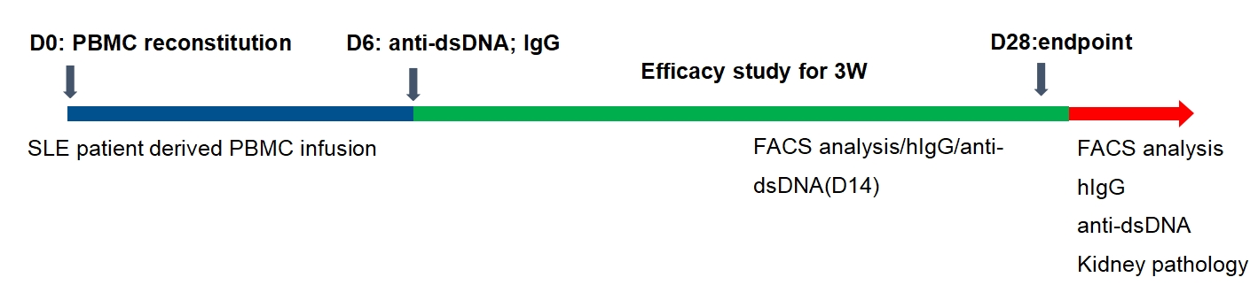
Immune Reconstitution SLE Models
Patient-derived PBMC-NCG SLE model
The levels of serum IgGs
D14 post reconstitution
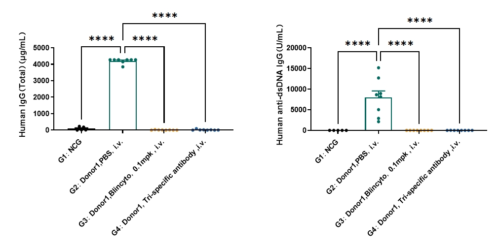
Treating with Blincyto and tri-specific antibody showed inhibition of human anti-dsDNA IgG in SLE model, as well as human total IgG one week after administration,
D28 post reconstitution
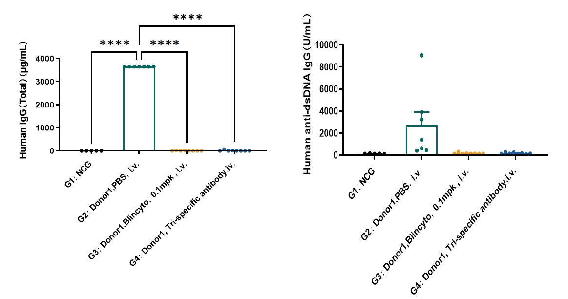
Blincyto and tri-specific antibody showed inhibition of human anti-dsDNA IgG in SLE model, as well as human IgG three weeks after administration.
Immune cells population
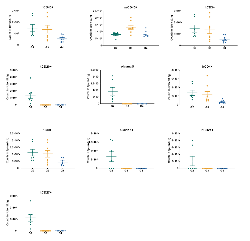
Blincyto and tri-specific antibody showed depletion of B cells (CD 20+), memory B cells (CD27+) and plasma B cells three weeks after administration.
IgG deposition in Kidney
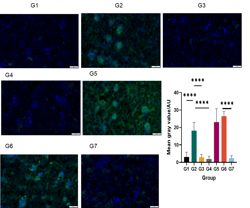
Blincyto and tri-specific antibody showed inhibition of human IgG deposition in SLE model three weeks after administration,
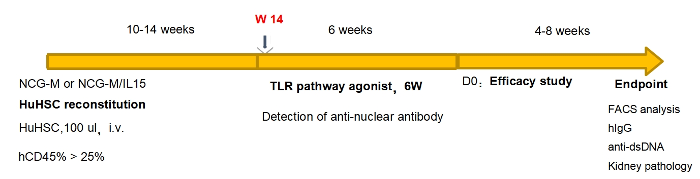
Induction of SLE model in HSC reconstitution mice
Serum IgG levels
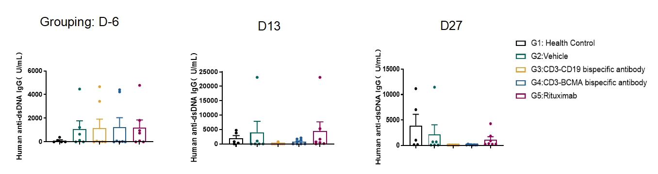
HSC-NCG SLE model responded to anti-CD3-CD19 bispecific antibody, anti-CD3-BCMA bispecific antibody and Rituximab with lower of serum anti-DNA IgG levels detected post-treatment.
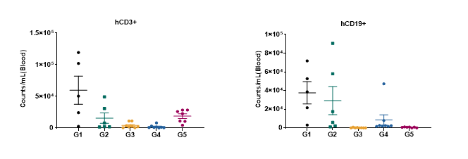
Immune cells population
D13 FACS of blood
D27 FACS of blood
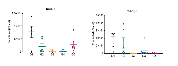
HSC-NCG SLE model responded to anti-CD3-CD19 bispecific antibody, anti-CD3-BCMA bispecific antibody and Rituximab with lower levels of circulating CD3+ and Cd19+ cells detected post-treatment.
D27 FACS of spleen
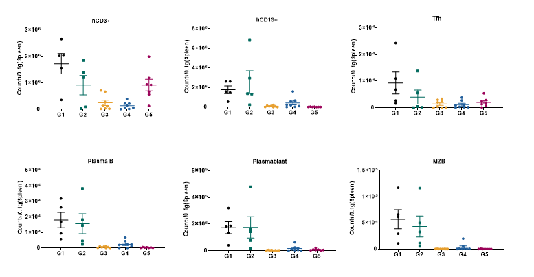
HSC-NCG SLE model responded to anti-CD3-CD19 bispecific antibody, anti-CD3-BCMA bispecific antibody and Rituximab with lower levels of CD3+, Cd19+, T follicular helper cells (TFH), plasma B-cells, plasmablast cells and marginal zone B-cells (MZB) detected post-treatment.