

Parkinson’s Disease Rat Model
Parkinson's disease (PD) is a progressive neurodegenerative disease that is characterized by a combination of motor and non-motor symptoms. GemPharmatech has successfully developed a PD rat model by precisely stereotaxically injecting 6-OHDA into both the Substantia Nigra pars compacta (SNc) and Ventral Tegmental Area (VTA). This well-constructed PD model offers a powerful tool for in-depth research and drug development, enabling a more comprehensive understanding and potential therapeutic interventions for PD.
6-OHDA induced PD rat model
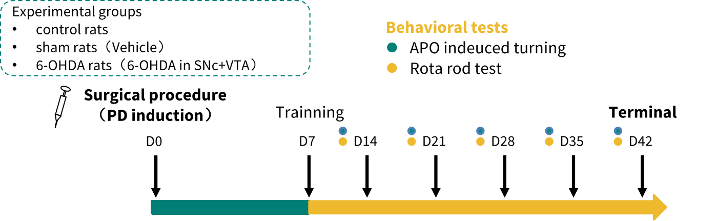
▎Model Construction Strategy
Data collection
Body weight
Survival proportion
Behavior test
Hematoxylin-eosin staining
Nissl body staining
Immunofluorescence staining (TH, Iba1, GFAP)
▎Body Weight Change and Survival Rate of the PD Model
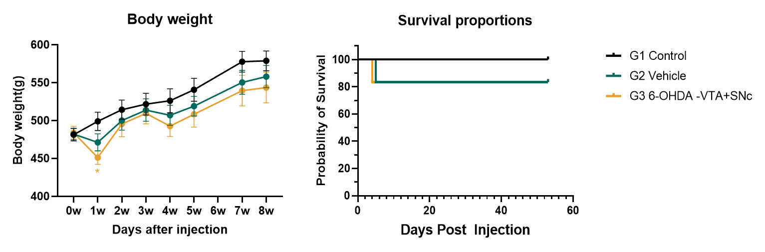
(data presented as Mean±SEM,n=6)
The body weight of the PD model was lower than the control group after unilateral stereotaxic injection. In the first week after unilateral stereotaxic injection of 6-OHDA, approximately 15% death was observed.
▎Confirmation of Successful Induction of PD by 6-OHDA
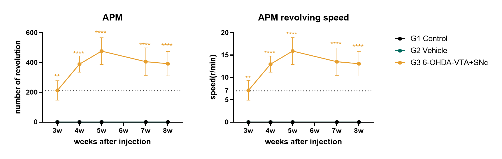
(data presented as Mean±SEM,n=6)
Three weeks after unilateral stereotaxic injection of 6-OHDA, subcutaneous injection of apomorphine (0.5 mg/kg) caused the rats to rotate toward the uninjured side. Rats that rotated more than 210 times (7 turns/minute) within 30 minutes were identified as PD models.
▎Behavior Test: Rotarod
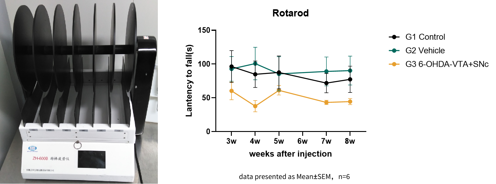
The PD rat model showed deficits in motor learning and motor coordination in the rotarod test.
▎Pathology: HE and NISSL body staining of Substantia Nigra
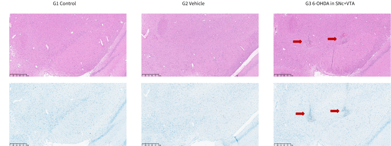
Unilateral stereotaxic injection of 6-OHDA into both the Substantia Nigra pars compacta (SNc) and Ventral Tegmental Area (VTA)
▎Pathology: IBA1+ and GFAP+ staining in Substantia Nigra
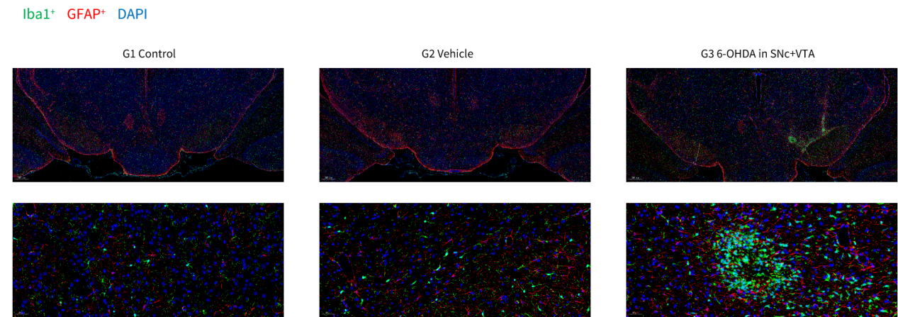
Eight weeks after 6-OHDA injection, IBA1+ (microglial marker) and GFAP+ (astrocyte marker) staining was significantly increased in the injection position of the substantia nigra of the PD model.
▎Pathology: TH+ neurons in Substantia Nigra and Striatum
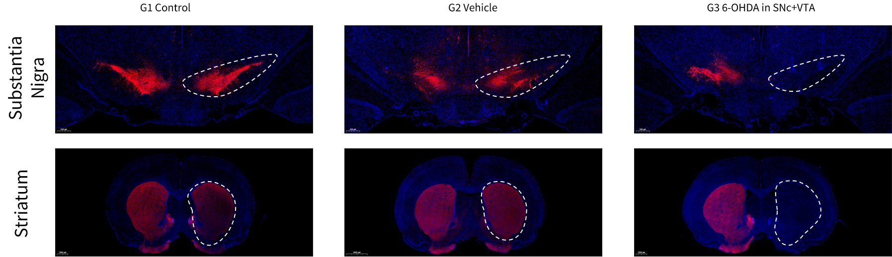
Eight weeks after 6-OHDA injection, TH+ neuron loss was observed in the striatum and substantia nigra on the injured side of the PD model.
▎Pathology: IBA1+ and GFAP+ staining in Striatum
Eight weeks after 6-OHDA injection, IBA1+ and GFAP+ staining in the striatum showed no difference among three groups.
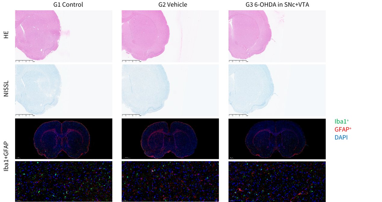
▎Pathology: IBA1+ and GFAP+ staining in Hippocampus
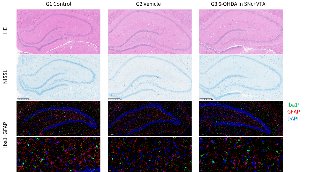
Eight weeks after 6-OHDA injection, IBA1+ and GFAP+ staining in the hippocampus showed no difference among three groups.